
The WAIT Test™ › Target Tissue
Target Tissue in Red Light Therapy: What It Means, Why It Matters, and How to Evaluate a Device
On this page
- Does the light reach the target tissue? The short answer
- Where Target Tissue fits in The WAIT Test™
- How to evaluate whether a device reaches the target tissue
- The WAIT Test™ standard for Target Tissue
- Common obstacles and how to overcome them
- Poor treatment advice and misleading product images
- Worked example: four devices on a hip
- The science: where light is lost on the way to the target tissue
- Technical details
- Delivery setups compared
- Air gaps, curves, and movement
- Contact at one point is not full-area contact
- Non-contact devices and distance
- Covers, films, lenses, diffusers, and spacers
- Lotions, gels, and skin preparation
- Skin pigment is different from fur pigment
- What equine studies add
- What is known across other species
- What about cats?
- Human hair
- Surface contact does not guarantee a deep dose
- What credible measurement looks like
- Evidence summary: what the research does and does not show
- What the evidence cannot currently answer
- Frequently asked questions
- What does “Target Tissue” mean?
- Does the device have to touch the skin?
- Does a wider beam really reach deeper?
- Can near-infrared light go through fur?
- Is touching my pet’s fur enough?
- Should I shave my dog or cat before treatment?
- Does dark fur absorb more light?
- Can red light therapy work through clothing?
- Can I use red light therapy over a bandage?
- Can the device touch an open wound?
- Does pressing harder help light go deeper?
- If a barrier cuts the light in half, can I double the time?
- Does a brighter-looking device deliver more light to the target?
- Does good contact prove the treatment will work?
- The bottom line
- Continue The WAIT Test™
- Research and references
For red light therapy to work, the light must reach the tissue that needs it and be absorbed by the cells there. That sounds obvious, but it’s where many treatments quietly fall short. Light that’s stopped by your pet’s fur, spread out by distance, or aimed at the wrong spot never gets the chance to help, no matter how good the device looks on paper. In red light therapy, also called photobiomodulation (PBM), getting the light all the way there is what we call reaching the target.
It’s also an often overlooked part of a good device. In our reviews of home devices, many companies don’t share even the basics: wavelengths, average power, and irradiance. Even companies with clear specifications often show their devices used from a distance or with a lip that keeps the light off the skin. Published bench tests found related problems: dose instructions that ignored beam spread and tissue losses (Cronshaw et al., 2025), and stated power and beam sizes that didn’t match measurements (Girasol et al., 2022).
Does the light reach the target tissue? The short answer
Only if three things are true. The device gets its light past the fur, it’s applied the way the condition needs (flush on bare skin for joints and the spine), and the company shows it being used that way.
Skin contact without air gaps is the most reliable way to get light to the target tissue. For pets, the coat is usually the biggest loss: The WAIT Test™ assumes only 25% of a device’s light gets through if the device rests on the fur instead of parting it.
Every Target Tissue evaluation asks the same question: does the light reach the target tissue?
Where Target Tissue fits in The WAIT Test™
Red light therapy has one job: get the right kind of light, in the right amount, to the target tissue. That’s why MedcoVet built The WAIT Test™.
The WAIT Test™ checks whether a red light therapy device can deliver a useful dose of light to the tissue that needs it. It has four components, and a device must pass all four:
Wavelengths
Is it the right kind of light for the target tissue?
The device must emit red or near-infrared light in a band that counts for the condition.Read the guide →Average Power
Is there enough light to treat the whole area?
The device’s true average optical output must cover the treatment area in one session.Read the guide →Irradiance
Is the light concentrated enough to deliver the dose in time?
The usable light at the skin must deliver the dose at each site within the time limit.Read the guide →Target Tissue
Does the light reach the target tissue?
The light must get past fur and be applied the way the condition needs.You’re reading this guideEach device is judged separately for each condition and coat, so the same device can pass for a wound and fail for a hip. The full method, including the data you need and the decision rules, is in The WAIT Test™ guide.
This guide covers the fourth component, Target Tissue. It’s the last link in the chain: the right light, in the right amount, still has to get to the tissue that needs it.
This guide starts with how to check whether a device’s light reaches the target tissue, including a worked example and the most common obstacles. Then it explains where light is lost between the device and the target, and it ends with technical detail.
How to evaluate whether a device reaches the target tissue
Ask three simple questions. They match the three Target Tissue checks in The WAIT Test™:
- Does it get through fur? (T1) Look for an applicator that parts the fur and reaches the skin, not one that rests on the coat.
- Is it applied the way the condition needs? (T2) For joints and the spine, that means sitting flush on bare skin with no air gap. If it’s used from a distance, the company should state the exact distance and how to keep it there.
- Do the company’s photos and videos show it used that way? (T3) A device shown held inches away, resting on the coat, or moved around can’t deliver the dose the company claims.
- Also worth checking: does the treatment area cover at least about 2 cm across? Wider areas let scattered light build up and reach deeper, but they need proportionally more total power to keep the same irradiance. This one isn’t a scored check.
- Bonus: can the company share a third-party report? A report showing irradiance measured at the finished device’s skin-facing surface adds confidence. It isn’t required, though. Plenty of good devices are made without one.
A device that checks all three boxes passes the Target Tissue checks for that condition. A third-party report is a plus, not a requirement.
Warning signs
- The device is shown resting on top of fur or clothing
- Product photos or videos showing the device held away from the skin or moved around during treatment
- No treatment distance is stated for a non-contact device
- Irradiance is measured at the LEDs, but the device is used from a distance
- Claims that light passes through fur or clothing, with no measurements
- Higher power offered as a substitute for reaching the skin
- Instructions to press hard to push light deeper
The standard
The WAIT Test™ standard for Target Tissue
The Target Tissue component has three checks, worth 25 points in all. Score each separately for every condition and coat.
- T1. Gets light through fur (5 points). For a coated pet, the device parts the fur so its light reaches the skin. If it sits on top of the fur, it fails T1, and the dose math in Average Power and Irradiance counts only 25% of its light.
- T2. Applied the way this condition needs (15 points). For joints and the spine, the device has to sit flush on the skin. A device that can’t sit flush on the skin fails. If the company doesn’t say whether it’s used in contact or at a set distance, T2 stays open.
- T3. Honest demonstrations (5 points). The company’s photos and videos show the device used as intended to deliver the dose, not held inches away, resting on the coat, or moved around. Until a device’s demonstrations have been assessed, T3 is left out rather than counted as a fail.
- Bonus: a third-party report showing irradiance measured at the skin-facing surface of the finished device adds confidence, but it isn’t required.
Target Tissue result
Pass or fail, per condition. T2 is worth 15 of the 25 points, so a device that can’t be applied the way the condition needs fails Target Tissue. T1 and T3 are worth 5 points each: missing one still passes, but missing both fails. A device that sits on the fur also loses most of its light in the Average Power and Irradiance dose checks, which is where fur usually costs a device its pass.
How The WAIT Test™ scores a device. Each device gets one result for each situation (the condition being treated and the coat over the area), not one result overall. Each check is worth a set number of points, and the checks are grouped under the four components. A device passes only if it clears the eye-safety gate and earns at least 75% of the available points in each of W, A, I, and T. It fails if any component stays below 75% even when every missing figure turns out as favorably as possible. If nothing has failed but a missing figure could still decide the result, it needs more information: it can’t pass until the figure is supplied, but it hasn’t failed either. The score out of 100 ranks devices and never overrides the result; when figures are missing, it’s shown as a range. Class 3B and 4 lasers and Risk Group 2–3 LEDs are not safe for home use and aren’t scored. See how every device does, condition by condition, in The WAIT Test™ Device Scorecard.
If the information isn’t available
Target Tissue is judged mostly from the device’s design, photos, videos, and instructions. If the company doesn’t say whether the device is used in contact or at a set distance, T2 stays open, and the device needs more information unless something else has already failed.
T3 (honest demonstrations) is left out of the available points until the device’s photos and videos have been reviewed, so a missing review neither helps nor hurts the device.
The one question to ask
Can you show how much light reaches the skin in the exact way I’m expected to use this device?
If the company can’t answer clearly, look at its photos, videos, and instructions. They decide T1, T2, and T3.
Common obstacles and how to overcome them
| Obstacle | Impact | How to overcome it |
|---|---|---|
| Fur | In 47 dogs, pooled median transmission was 0.9% (white coats), 0.3% (brown), and 0% (black), though individual readings reached 12–19% (Hochman-Elam et al., 2020) | Part the fur, or use an applicator that reaches the skin. Clip only with veterinary guidance. |
| Distance | From 5 cm away, no light was detected through skin and muscle in dogs (Kampa et al., 2020) | Use the device in contact with bare skin. For a non-contact device, use the exact stated distance. |
| Angle, curves, and air gaps | Light spreads thin, and the edges of the pad get less | Use a pad that sits flush, and hold it straight against the skin. |
| Clothing and blankets | One layer of cotton can block about two-thirds of near-infrared light (Dubrovski et al., 2023) | Treat bare skin. Don’t treat through clothing or blankets. |
| Bandages and dressings | Some gels and membranes let more than 50% through; others block most of it (Lilge et al., 2000) | Treat through a dressing only if your veterinarian approves and it has been tested. |
| Covers, films, and lenses | Can absorb or redirect light and change the wavelength mix | Make sure the company measured the device with them in place. |
| Lotions and gels | Can reflect, scatter, or absorb light | Treat clean, dry skin unless the device is designed for a gel. |
| Reflection at the skin | Some light is lost at the surface, more with an air gap | Keep the device flush against the skin. |
| Narrow beam | Light scatters out the sides and doesn’t go as deep | Choose a device that covers a wider treatment area. |
| Deep target | Through dog skin, about 8 to 9% got through; through skin and muscle, less than 1% (Kampa et al., 2020) | Combine contact, gentle pressure, a wide beam, and near-infrared light. |
| Movement | Each spot gets a different amount of light | Use a strap or hold the device still for the full session. |
| Wrong spot | The light never reaches the tissue that needs it | Follow clear placement instructions or your veterinarian’s guidance. |
Fur: what the dog studies show
For pets, fur is usually the biggest obstacle. Touching the coat is not the same as reaching the skin.
A study of 47 healthy dogs tested two veterinary lasers on black, brown, and white coats and measured the light that passed through. Laser power, wavelength, coat color, and shaving all had a significant effect. Darker coats let through less light, and shaved sites let through more. Coat length wasn’t a significant factor in the study’s statistical model (Hochman-Elam et al., 2020).
This doesn’t give one loss percentage for every dog, and it doesn’t mean coat length never matters. Coat density, undercoat, hair thickness, skin pigment, wavelength, and how well the applicator parts the fur all interact. And turning up the power isn’t a substitute for getting the light to the skin.
How The WAIT Test™ accounts for fur: if a device doesn’t part the fur, the test assumes only 25% of its light gets through the coat. A dark (brown) coat, assumed to mean darker skin too, passes 33% as much light as a light coat. Black coats pass less still and aren’t scored separately. Shaved areas and clipped wounds are exempt from these losses. Coat length isn’t used, in line with the 47-dog study.
Does fur need to be shaved?
Not always. The goal is a repeatable path to the skin. Depending on the pet and the device, that might mean:
- Parting the fur by hand
- Using an applicator that slips between the hairs
- Using a comb-style applicator that separates the coat
- Clipping a small area when your veterinarian thinks it’s appropriate
- Using an area already clipped for surgery, following post-op instructions
Shaving removes the most hair, but it isn’t generally recommended. It can irritate skin, disturb a double coat, and stress the animal. Dark coats also absorb more light at the surface, which can warm the skin. In the 47-dog study, the authors recommended shaving dark-coated dogs and monitoring heat, since black-coated areas showed skin reactions at the highest power setting (Hochman-Elam et al., 2020). Don’t shave an injured, infected, inflamed, or recently operated area without veterinary advice.
What a pet device should show
- Diagrams or photos showing the applicator parting the fur and touching the skin
- Irradiance measured at the skin-facing surface of the finished device
- Testing across different coat colors and densities
- Clear instructions on when clipping is needed
- Clear limits on coats where the device can’t reliably reach the skin
A claim that “near-infrared penetrates fur” isn’t enough. Some light may get through a coat, but how much, which wavelengths, and where it lands may be unknown.
Comb-style applicators
A comb can solve the fur problem by separating the hair and placing the light close to the skin. Human laser combs tested in randomized, sham-controlled trials are one example of this design (Jimenez et al., 2014). But scalp results don’t prove a pet comb works. Judge a pet comb by whether its teeth reach the skin safely, whether the light comes out close to the skin, and whether the whole device was measured that way.
Clothing
Clothing adds another barrier to an already hard path. By the time light reaches a joint or tendon, the skin, fat, and muscle have already absorbed most of it. Clothing takes its share before the light even reaches the skin.
And that share is large. In a 2023 study, a single layer of woven cotton let through only about 26 to 37% of near-infrared light at 950 nm, so roughly two-thirds or more was blocked. Darker dyes blocked even more at shorter wavelengths (Dubrovski et al., 2023). Clothing doesn’t stop all near-infrared light (Jeffery et al., 2025), but some light getting through isn’t the same as a useful dose. Laser therapy experts Tunér, Hode, and Jenkins put it simply: clothes are “a very effective blocker of light” (Tunér, Hode, and Jenkins).
The fix is easy: treat bare skin. Removing clothing is one of the simplest ways to give light a real chance to reach the target.
Bandages, wounds, and fragile skin
A wound dressing may be medically necessary. Don’t remove it just to let more light through unless your veterinarian says it’s safe. Dressings vary widely: some gel and membrane dressings let more than 50% of the light through in laboratory testing, but you must measure any untested product at the intended wavelength (Lilge et al., 2000).
Direct contact may be inappropriate over open wounds, recent incisions, infected areas, burns, or painful or fragile skin. A spacer or non-contact method can keep the device away from the tissue, but the spacer becomes part of the light path and should be included in the measurements. PBM should never delay diagnosis or replace wound care, antibiotics, surgery, or pain management.
Poor treatment advice and misleading product images
Sometimes the biggest obstacle isn’t the device. It’s the instructions. Even a well-designed device will miss the target when owners are told to:
- Rest the device on top of the coat instead of parting the fur and reaching the skin
- Treat the wrong spot, such as where the pet licks rather than over the joint or tissue causing the problem
- Hold a panel “a few inches away” without a stated distance or a way to keep it there
- Move the device around without a defined path or time on each spot
- Treat through clothing or a blanket without measurements that support it
- Press hard to push light deeper, which can cause discomfort without a proven benefit
Good instructions tell you exactly where to place the device, how to get it to the skin, how long to hold it still, and what to do differently over a wound or surgical site.
Misleading product images
Watch how a company shows its own device being used. Photos and videos of a device held inches from the skin, resting on the coat, or moved around during treatment aren’t just a styling choice. They’re misleading. A device used that way can’t deliver the dose the company claims, because distance, fur, and movement waste most of the light before it reaches the target tissue.
It also raises a bigger question. If a company’s own images don’t match how its device needs to be used, can you trust its dosage numbers, treatment times, and other materials? Treat that mismatch as a red flag for everything else it publishes.
Worked example
Worked example: four devices on a hip
Here’s how the Target Tissue checks play out for four devices in The WAIT Test™ device comparison, for a 50 lb dog’s hip under a light coat.
| Device | Through fur (T1) | Flush on the skin (T2) | Honest demonstrations (T3) | Target Tissue | Knock-on effect |
|---|---|---|---|---|---|
| MedcoVet Luma | Parts the fur | Yes | Yes | 100%: pass | All of its light counts in the dose checks |
| B-Cure Laser Vet Pro | Sits on the fur | Yes | Yes | 80%: pass | Only 25% of its light counts, so it fails Average Power and Irradiance |
| LumaCare Ark | Sits on the fur | Yes | Misleading | 60%: fail | Two smaller misses together fail the component |
| Whisker Bark Red Light Therapy Device | Sits on the fur | Not held flush | Not yet assessed | 0%: fail | Can’t be applied the way a hip needs |
The B-Cure shows why Target Tissue isn’t the only place the coat matters. It passes this component, but resting on the fur cuts its usable light to a quarter, and that’s where it fails. Target Tissue checks how the device is applied; Average Power and Irradiance check whether enough light survives the trip.
Deep dive
The science: where light is lost on the way to the target tissue
What can prevent light from reaching the target?
A device can have the right wavelengths, an honest average power, and a good irradiance, and still lose most of its light before it reaches the tissue. Light can be lost in two places: on the way to the skin, and on the way through the body.
| What gets in the way | Where | What it does |
|---|---|---|
| Fur and hair | Before the skin | Absorbs and scatters light. Darker and denser coats let less through. |
| Distance | Before the skin | Light spreads out, and much of it misses the body. |
| Angle and air gaps | Before the skin | A tilted device, or a pad that doesn’t sit flush over a curved joint, spreads the light thin and leaves weak spots. |
| Clothing, blankets, and bandages | Before the skin | Block, scatter, or filter light, sometimes more of one wavelength than another. |
| Covers, lenses, and lotions | Before the skin | Absorb or redirect light before it reaches the skin. |
| Poor treatment advice | Before the skin | The device rests on the coat, sits on the wrong spot, or is moved around. |
| Reflection at the skin | At the skin | Some light bounces off the surface, more so when there’s an air gap. |
| The tissue itself | Inside the body | Skin, fat, blood, and muscle absorb and scatter light. The deeper the target, the more is lost. |
| A narrow beam | Inside the body | Light scatters out the sides, so it doesn’t go as deep. |
You can’t change the tissue, but you can control almost everything else. The goal is to remove as many of these losses as possible. What matters isn’t how much light leaves the device. It’s how much reaches the target tissue and is absorbed there.
So why is reaching the target hard?
Even though cells are tiny and only a little light is needed, almost everything between the device and the target either reflects or absorbs it. A few numbers give a sense of scale:
- Skin: in human skin, 850 nm laser light lost 66% of its intensity in less than 1 mm through a 0.78 mm ex vivo sample with a very narrow beam. (Esnouf et al., 2007).
- Skin and muscle: in living dogs, only about 8 to 9% of 830 nm laser light applied in contact made it through the skin, and just 0.25 to 0.78% made it through skin and muscle (Kampa et al., 2020).
- Distance: with the probe 5 cm away, no light was detected through skin and muscle at all (Kampa et al., 2020).
- Fur: in 47 dogs, the pooled median transmission (across two lasers, several power settings, and two test sites) was 0.9% for white coats, 0.3% for brown coats, and 0% for black, though individual readings reached 12% to 19% in every color. Shaved sites had a higher median than unshaved sites (0.7% vs. 0%) (Hochman-Elam et al., 2020).
- Bone: in sectioned, preserved cadaver tissue, roughly 1% to 12% of 830 nm light made it through the scalp and skull, depending on the site (Jagdeo et al., 2012).
Each loss multiplies the others. Put fur, a gap, and a deep target together, and what’s left can be too little to help. That’s why a device needs far more light at the skin than the target does: the extra light at the surface covers the losses on the way down. The WAIT Test™ sets the minimum at the target at 5 mW/cm², then divides by the share of light that reaches the target tissue’s depth. That works out to about 11 mW/cm² of usable light at the skin for most joints and the spine, 17 for the shoulder, and 25 for the hip, counted after the coat takes its share.
We’ll cover each of these in more detail below, along with how to avoid them.
Understanding the path between the device and the target
Light’s path to the target has two parts:
- Before it reaches the skin. Distance, angle, air gaps, fur, clothing, and covers all act here.
- Once it enters the skin, it travels through everything between the skin and the target: skin, fat, blood, muscle, and tendon.
You can improve both parts of the path, though many companies ignore them. The first part decides how much light enters the body. The second determines how much reaches the target tissue.
Part 1: Before the light reaches the skin
Everything between the device and the skin can waste light before it ever enters the body:
- Distance: as the device moves away from the skin, the beam spreads, and much of the light misses the body. Small shifts change the beam size, irradiance, and covered area.
- Angle: a tilted device spreads its light over a larger, stretched area, so less power lands on each square centimeter.
- Air gaps and curves: a rigid pad can touch a joint at one point while its edges float above the skin, leaving weak spots.
- Fur and hair: a coat absorbs and scatters light before it reaches the skin. Dark, dense coats block the most.
- Clothing, blankets, and bandages: each can block, scatter, or filter light.
- Covers, lenses, films, and lotions: anything between the light source and the skin can absorb or redirect light.
- Movement: when the device slides around or your pet shifts, each spot gets a different amount of light.
How to improve the path to the skin
- Put the device in direct contact with bare skin, with no air gap. This removes distance and angle losses in one step.
- Get through the fur. Part the coat, or use an applicator designed to slip between the hairs and reach the skin.
- Use a pad that fits the body. A flexible or well-shaped pad sits flush over curves instead of touching at one point.
- Hold it still. Use a strap or steady hand to keep the device in place for the full session.
- Treat bare skin. Remove clothing and blankets, and only treat through a dressing if your veterinarian approves and it has been tested.
- Check the measurements. Measure irradiance with any covers or lenses in place, at the skin-facing surface.
Skin contact makes it easier to repeat placement. It establishes and maintains distance, prevents unintended angling, and can reduce losses from fur or surface reflections. Furthermore, some experiments involving tissue transmission have found greater delivery with contact than without contact. However, contact alone does not prove the target received an effective dose, and it does not mean the other WAIT criteria can be ignored.
Even companies that mainly sell non-contact panels acknowledge this. For example, GembaRed writes that “the promise of deep penetration is only delivered with skin-contact devices” (GembaRed).
The dog studies show how much this matters. In dog cadavers, applying 980 nm light in contact increased the light detected inside the spinal canal by as much as 67% compared with applying it off contact (Piao et al., 2019). In living dogs, 830 nm light applied in contact was still measurable through skin and muscle, while the same light from 5 cm away wasn’t detected (Kampa et al., 2020).
Part 2: From the skin to the target
Reflection at the skin
At the air-skin interface, some light is reflected because the two materials have different refractive indices. A contact interface can affect this boundary, and it can also push hair or fur aside so the beam doesn’t have time to spread before it reaches the body. To put that in perspective, only about 3 to 4% of the light bounces straight off the surface, because skin’s refractive index (about 1.4 to 1.5) isn’t much higher than air’s (1.0). Much more light enters the skin, scatters, and returns. That same scattering is what makes the light just below the surface brighter than the light arriving at it (Jacques, 2010).
This is another reason skin contact without an air gap matters. With the device flush against the skin, there’s no gap for light to escape, and light that bounces back at the surface is more likely to be sent back in. The exact gain depends on the applicator, wavelength, pressure, skin, and anatomy, so measure it rather than assign one universal percentage.
Inside the body
Once light enters the skin, melanin, blood, water, and fat absorb and scatter it, and the deeper the target, the smaller the share that arrives. The Wavelengths guide explains why near-infrared travels farther than red. The WAIT Test™ turns this into one number for each condition: the share of light that reaches the target’s depth, from 45% for most joints and the spine down to 20% for a hip.
A wider beam reaches deeper
Most light in tissue doesn’t travel in a straight line. It scatters, bouncing off cells and fibers in every direction. That scattering has a surprising effect: just under the skin, light builds up into a bright zone that can be 3 to 5 times brighter than the light arriving at the surface (Jacques, 2010).
Beam size decides how much of that scattered light ends up hitting the target:
- With a narrow beam, sideways-scattered light leaves the beam and isn’t replaced. The center fades sooner, and less light reaches deep tissue.
- With a wide beam, light from the surrounding area scatters into the target from every side. Light that scatters out of one spot is replaced by light scattering in from its neighbors, so more of it builds up and travels deeper.
Computer models of light in tissue show this clearly: as the beam diameter shrinks, the light at the beam center drops off faster with depth (Keijzer et al., 1989). Beam edges lose light, so a beam needs to be about six penetration depths wide before it behaves like a “broad” beam (Jacques, 2010). As one PBM editorial put it, light dosing and tissue penetration are complicated, and spot size is one factor that changes them (Lanzafame, 2020).
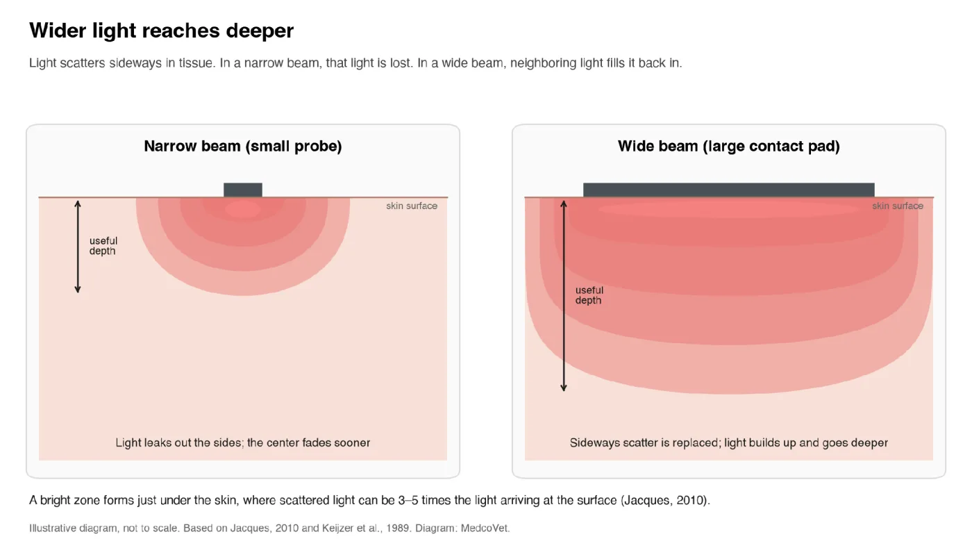
Figure 1. At the same irradiance, a wide contact pad builds up more light under the skin and carries it deeper than a small probe. Illustrative, based on Jacques (2010) and Keijzer et al. (1989). Diagram: MedcoVet.
At the same irradiance, a device that covers a wider skin area will generally deliver more light to deeper tissue than a small probe touching a single spot.
Gentle pressure can improve penetration
Contact and pressure are not the same thing. A device can rest lightly on skin, or it can compress the tissue.
Low mechanical compression can thin the tissue layer, shift blood and water, and change scattering. Optical studies have shown that light penetration increases with compression. In a study of the human forearm using optical coherence tomography, pressures between 4 and 8 kPa reduced the dermal optical attenuation coefficient by at least 1.0 mm⁻¹ (Bachour et al., 2023). Pin-array devices pressed into the skin improved light penetration in living human skin and in ex vivo pig skin and fat (Drew et al., 2009; Rylander et al., 2008).
More studies point the same way. In pig skin, pressing on the tissue increased light transmission and reduced scattering by about 12% at strong compression and about 36% at very strong compression (Vogt et al., 2012). Compression also pushed water out of the skin, which reduces scattering (Gurjarpadhye et al., 2011). A compression-controlled probe used negative pressure to concentrate laser light in pig skin, raising peak intensity by about 2.7 to 3.6 times (Yeo et al., 2010). An earlier study found that compression changes soft tissue optical properties (Chan et al., 1996).
These were optical studies, not clinical trials showing that more pressure improves PBM results. Laser therapy experts Tunér, Hode, and Jenkins also note that a lower-powered laser “applied in contact and with slight pressure to the skin” may penetrate more deeply than a high-power laser. The practical lesson: use gentle, consistent contact as the device instructions describe. Gentle, steady pressure helps, but pressing hard doesn’t add more and can hurt. Avoid pressure that causes pain or presses on fragile tissue.
Putting it together: skin contact without air gaps
Skin contact without air gaps improves both parts of the path. Before the skin, it removes distance, angle, and most fur losses. At the skin, it reduces surface light loss. Below the skin, it enables gentle pressure, and a wide contact pad lets scattered light build up and reach deeper. That’s why skin contact is the most reliable way to reach the target.
Technical reference
Technical details
The sections below go deeper into the technical side of reaching the target. You don’t need them to compare devices, but they’re useful if you want to understand the details.
Delivery setups compared
Researchers describe three broad delivery modes: non-contact, contact, and deep (light delivered inside the body). They note that contact delivery suits treatments that need to concentrate light in one area (Rojas and Gonzalez-Lima, 2011).
There are four common delivery setups:
| Delivery setup | Likely to reach the target? | What must be specified |
|---|---|---|
| Contact on uncovered skin | Yes, most reliably | Contact area, how it stays in place, irradiance at the skin, and any lens or cover in the path |
| Non-contact over uncovered skin | Yes, if distance and angle are controlled | Working distance, angle, illuminated area, irradiance at the skin, and a way to hold the position |
| Through a required barrier, such as a dressing | Sometimes | The exact barrier, how much light it lets through, irradiance after the barrier, and an adjusted protocol |
| Resting on fur, clothing, or at an uncontrolled distance | Not reliably | Actual distance, angle, coverage, and how much light gets through are unknown |
Air gaps, curves, and movement
Air doesn’t absorb much red or near-infrared light over normal treatment distances. The problem with an air gap is what happens to the beam while crossing it: distance, angle, overlap, and coverage all change. On a curved joint, the center of a rigid applicator may be close to the skin while the edges are farther away and angled, so the exposure drops from center to edge even when every LED is working.
Movement adds another variable. Scanning can be a valid professional technique, but it needs a defined speed, path, overlap, and number of passes. Accidental movement isn’t a controlled scan.
Contact at one point is not full-area contact
A pad may feel secure while parts of it float above the body: a flat pad bridging the sides of a knee, a wrap lifting at the edges, a rigid head rocking over a hip, or a pet applicator sitting on guard hairs. The treatment area is the part that actually receives the intended irradiance, not the product’s outer size.
Non-contact devices and distance
Panels and lamps can provide a controlled treatment when the geometry is clear. A complete non-contact protocol should state the distance, allowed tolerance, angle, illuminated area, area-average irradiance at the skin, light uniformity, and how to maintain position.
As distance changes, irradiance, beam size, LED overlap, edge falloff, and the amount of light that misses the body all change together. The inverse-square law is often applied as if a panel were a single point of light. A nearby LED array is a large source with many overlapping beams, so a simple inverse-square calculation can be a poor substitute for measuring at the actual treatment distance.
Covers, films, lenses, diffusers, and spacers
Accessories may be needed for hygiene, comfort, or distance, but they’re also part of the light path. Each can change the total power, the wavelength mix, the beam width, hot and dark spots, and heat at the skin. If a device has two approved setups, such as direct contact and a wound spacer, each needs its own distance, irradiance, area, and treatment time.
Lotions, gels, and skin preparation
Topical products contain pigments, particles, oils, water, and polymers that can affect reflection, scattering, and absorption. Something that looks clear isn’t necessarily clear to red and near-infrared light. Follow the device instructions for clean, dry skin, don’t add coupling gel unless the device was designed and tested for it, and don’t use alcohol to let more light through.
Skin pigment is different from fur pigment
Removing hair doesn’t remove the melanin in the skin. Skin pigment affects absorption and surface heating, while hair and fur add their own absorption and scattering. For pets, consider coat color and skin color separately: a light coat can hide dark skin, and a dark coat can hide lighter skin.
What equine studies add
In nine horses, researchers applied an 810 nm, 500 mW probe to 17 forelimbs and measured light on the far side of the flexor tendon. Clipping significantly increased light transmission. Cleaning with alcohol without clipping didn’t make a significant difference (Ryan and Smith, 2007).
An ex vivo equine study of 800 and 970 nm light also found that hair, skin, preparation, wavelength, and tissue thickness affected how much light passed through skin and tendon (Duesterdieck-Zellmer et al., 2016). The alcohol result isn’t advice for home use: alcohol can irritate skin and isn’t appropriate on wounds.
What is known across other species
A 2024 postmortem study tested thin skin samples from species with fur, feathers, scales, and different pigmentation. Lighter samples generally let more light through than darker ones, and feathers let little or no light through (Ramsey et al., 2024). The authors noted that studies in living animals are still needed.
What about cats?
We didn’t find a cat study comparable to the dog studies above. Cats differ from dogs in coat, skin, size, behavior, and common treatment sites, so don’t apply dog numbers to cats. For cats, use a device that reaches the skin, avoid relying on product photos that show light resting on the coat, ask your veterinarian before clipping, and stop if your cat becomes stressed or the area feels too warm.
Human hair
Hair can block, scatter, and redirect light before it reaches the scalp. For a scalp device, ask whether it separates the hair or just shines on top of it, and whether irradiance was measured at the scalp.
Surface contact does not guarantee a deep dose
The dose at the skin is:
Surface radiant exposure (J/cm²) = irradiance at the skin (W/cm²) × time (s)
That doesn’t account for losses in a barrier, reflection at the skin, scattering and absorption in tissue, tissue thickness, or the distance to the target. If a barrier lets through a measured fraction of light, a first estimate is:
After-barrier irradiance = bare-path irradiance × barrier transmittance
For example, if 50 mW/cm² hits a material that lets through 40%, about 20 mW/cm² comes out the other side, before accounting for any change in the beam pattern. This only works when the transmittance was measured at the same wavelength, geometry, and material condition.
What credible measurement looks like
PBM reporting guidance lists the beam area at the skin as a key parameter and asks researchers to report the application technique, including contact, projection, scanning, and pressure (Jenkins and Carroll, 2011). A systematic review of 74 PBM papers found that 73% didn’t describe how they measured light, 43% didn’t report irradiance, and 40% didn’t report beam area (Hadis et al., 2016).
Recent bench tests of five home-use LED PBM devices found considerable beam spread and concluded that the manufacturers’ dose instructions didn’t consistently account for beam spread and tissue attenuation (Cronshaw et al., 2025).
Evidence summary: what the research does and does not show
| Study | Model | Finding | Limitation |
|---|---|---|---|
| Karu et al., 2004 | HeLa cells (a human cell line), 600–860 nm | Cells attached better after about 0.005 J/cm² | Lab dish, not living tissue |
| Kampa et al., 2020 | 24 living dogs, 830 nm, skin and muscle | 8–9% through skin in contact; 0.25–0.78% through skin and muscle; none detected at 5 cm | Specific 200 mW probe and post-surgical model |
| Hochman-Elam et al., 2020 | 47 living dogs, two veterinary lasers | Coat color, shaving, wavelength, and power affected transmission | No universal coat correction factor |
| Piao et al., 2019 | Six dog cadavers, 980 nm, spinal canal probe | Contact increased detected light by up to 67% versus off-contact | Cadaver model, specific wavelength and anatomy |
| Esnouf et al., 2007 | Human skin samples, 850 nm | 66% of the light lost in less than 1 mm | Thin ex vivo samples, narrow beam |
| Jagdeo et al., 2012 | Sectioned, preserved cadaver tissue, 830 nm | Roughly 1–12% through scalp and skull, depending on site | Preserved cadaver tissue |
| Ryan and Smith, 2007 | Nine horses, 17 limbs, 810 nm | Clipping increased transmission; alcohol alone did not | Small sample, one anatomical path |
| Ramsey et al., 2024 | Postmortem samples from many species | Pigment and coverings, especially feathers, reduced detected light | Thin frozen-thawed samples |
| Jacques, 2010 | Tissue optics review and modeling | Light just under the skin can be 3–5× the incident light; narrow beams lose light at the edges | Modeling, not a clinical outcome |
| Keijzer et al., 1989 | Monte Carlo model of finite-diameter beams | Smaller beams lose light faster with depth | Modeled artery tissue |
| Tedford et al., 2015 | Human cadaver heads, 808 nm | Near-infrared light detected about 40 mm deep | Cadaver tissue, one wavelength |
| Bachour et al., 2023 | Human forearm skin measured by OCT | Low compression reduced optical attenuation | Optical result, not PBM efficacy |
| Lilge et al., 2000 | Laboratory testing of wound dressings | Some gels and membranes let through more than 50% | Product- and wavelength-specific |
What the evidence cannot currently answer
Research doesn’t yet provide a single contact-advantage percentage for all devices, a universal loss factor for clothing or fur, a conversion from coat color to treatment time, a cat-specific coat table, or proof that more light at the target always improves clinical outcomes. Veterinary PBM research also has wider limits: a 2023 systematic review of 45 studies in dogs, horses, and one cat found wide variation in methods and risk of bias (Millis and Bergh, 2023).
FAQ
Frequently asked questions
What does “Target Tissue” mean?
It’s the T in The WAIT Test™. It checks whether a device gets its light through fur, is applied as the condition requires (flush on the skin for joints and the spine), and is shown being used honestly. Fur, distance, angle, barriers, and placement all determine how much light reaches the tissue you want to treat.
Does the device have to touch the skin?
No, but contact is the most reliable option. A non-contact device needs a stated working distance, a way to hold that distance, and irradiance measured at the skin at that distance.
Does a wider beam really reach deeper?
Yes, at the same irradiance. Light scatters sideways in tissue. With a wide beam, neighboring light fills in what scatters away, so light builds up and travels deeper. With a narrow beam, that sideways light is lost (Jacques, 2010; Keijzer et al., 1989).
Can near-infrared light go through fur?
Some can, but “some” isn’t a dose. Studies in dogs and horses show that coat color and coat preparation change how much light gets through. A pet device should part the fur and reach the skin, or show measurements through representative coats.
Is touching my pet’s fur enough?
No. The surface that matters is the skin. A device resting on the coat can leave an air gap and lose light in the hair before it reaches the body.
Should I shave my dog or cat before treatment?
Not without a reason and veterinary guidance. Parting the fur or using an applicator that reaches the skin may be enough. Clipping can let more light through, but shaving can irritate skin and isn’t right for every animal, coat, or wound.
Does dark fur absorb more light?
In a study of 47 dogs, darker coats were linked to less light getting through. That result doesn’t give a universal correction factor, because hair density, skin pigment, wavelength, and device design also matter.
Can red light therapy work through clothing?
Not well. Clothing adds another barrier to an already difficult path. A single layer of cotton can block about two-thirds of near-infrared light, and the skin and tissue take much of what’s left. Some light may get through, but not reliably enough to count on. Treat bare skin.
Can I use red light therapy over a bandage?
Only if your clinician allows it and the exact dressing has been evaluated. Some gels and membranes let enough light through, while other dressings block or scatter most of it.
Can the device touch an open wound?
Don’t assume so. Direct contact can cause pain, damage, or contamination. A spacer or non-contact method may be appropriate, but it needs its own measured irradiance and protocol.
Does pressing harder help light go deeper?
Compression can change how light travels through tissue, but no clinical rule says more pressure yields better PBM results. Too much pressure can cause pain. Use the gentle, defined contact the instructions describe.
If a barrier cuts the light in half, can I double the time?
Not automatically. A barrier can also change the beam pattern, the wavelength mix, and heat. Because PBM doesn’t reliably follow reciprocity, lower irradiance for longer doesn’t produce the same effect.
Does a brighter-looking device deliver more light to the target?
Not necessarily. Your eye isn’t a light meter, and it can’t see near-infrared light at all. Visible brightness tells you nothing about the irradiance at the skin or how much light reaches the target tissue.
Does good contact prove the treatment will work?
No. It makes the starting conditions reliable. The result still depends on wavelength, power, irradiance, time, schedule, the target, the condition, and the individual animal.
The bottom line
Reaching the target means getting light to the tissue that needs it, not just pointing it in the right direction. Cells need surprisingly little light, but fur, distance, air gaps, the skin, and the tissue below can take almost all of it.
Skin contact without air gaps is the most reliable way to protect that light. It fixes distance, controls the angle, moves fur out of the way, and reduces surface light loss. A wider contact area helps too, because a wide beam carries light deeper than a narrow one.
Before choosing a red light therapy device, ask:
Can the company show how much light reaches the skin in the exact way I’m expected to use the device?
If the company can’t connect its numbers to that real treatment setup, the T portion of The WAIT Test™ is incomplete.
Continue The WAIT Test™
This guide covers Target Tissue, the fourth of the four components. To run the full evaluation, including the data you need and the decision rules, use The WAIT Test™ guide. The other three components:
- Wavelengths: Is it the right kind of light for the target tissue?
- Average Power: Is there enough light to treat the whole area?
- Irradiance: Is the light concentrated enough to deliver the dose in time?
The four components work together. Passing one does not compensate for missing information in another.
The WAIT Test™ was developed by MedcoVet. The framework may be shared for educational purposes when its four components remain unchanged and MedcoVet is credited as its creator.
The WAIT Test™ is an educational framework for comparing device specifications. It does not determine whether PBM is appropriate for a specific pet, diagnosis, wound, or treatment area. Make treatment decisions with a qualified veterinary professional.
For every WAIT guide and tool in one place, start at The WAIT Test™ hub.
Ready to take the next step?
Book a free consultation with a licensed clinician to discuss your pet’s condition and treatment plan.
Hazel Moon
Learn more about the MedcoVet Luma and how its contact applicator is designed to move fur out of the light path for at-home treatment.
Sources
Research and references
- Bachour RPS, Dominguez CT, Cardoso GC. Optical attenuation coefficient of skin under low compression. Journal of the Optical Society of America A. 2023;40(5):955-960. PubMed
- Chan EK, Sorg B, Protsenko D, O’Neil M, Motamedi M, Welch AJ. Effects of compression on soft tissue optical properties. IEEE Journal of Selected Topics in Quantum Electronics. 1996;2(4):943-950. DOI
- Chung H, Dai T, Sharma SK, Huang YY, Carroll JD, Hamblin MR. The nuts and bolts of low-level laser (light) therapy. Annals of Biomedical Engineering. 2012;40(2):516-533. Full text
- Cronshaw M, Parker S, Hamadah O, Arnabat-Dominguez J, Grootveld M. Photobiomodulation LED devices for home use: design, function, and potential: a pilot study. Dentistry Journal. 2025;13(2):76. PubMed
- Drew C, Milner TE, Rylander CG. Mechanical tissue optical clearing devices: evaluation of enhanced light penetration in skin using optical coherence tomography. Journal of Biomedical Optics. 2009;14(6):064019. PubMed
- Dobnik Dubrovski P, Fakin D, Ojstršek A. Cotton woven fabrics as protective polymer materials against solar radiation in the range of 210-1200 nm. Polymers. 2023;15(5):1310. Full text
- Duesterdieck-Zellmer KF, Larson MK, Plant TK, Sundholm-Tepper A, Payton ME. Ex vivo penetration of low-level laser light through equine skin and flexor tendons. American Journal of Veterinary Research. 2016;77(9):991-999. PubMed
- Esnouf A, Wright PA, Moore JC, Ahmed S. Depth of penetration of an 850nm wavelength low level laser in human skin. Acupuncture & Electro-Therapeutics Research. 2007;32(1-2):81-86. PubMed
- GembaRed. 810nm vs 830nm: the best wavelengths for red light therapy. Company blog. Website
- Girasol CE, Braz GA, Bachmann L, Celli J, Guirro RRJ. Laser light sources for photobiomodulation: the role of power and beam characterization in treatment accuracy and reliability. PLOS ONE. 2022;17(3). Full text
- Gurjarpadhye AA, Vogt WC, Liu Y, Rylander CG. Effect of localized mechanical indentation on skin water content evaluated using OCT. International Journal of Biomedical Imaging. 2011;2011:817250. PubMed
- Hadis MA, et al. The dark art of light measurement: accurate radiometry for low-level light therapy. Lasers in Medical Science. 2016;31(4):789-809. PubMed
- Haslerud S, et al. Achilles tendon penetration for continuous 810 nm and superpulsed 904 nm lasers before and after ice application: an in situ study on healthy young adults. Photomedicine and Laser Surgery. 2017;35(10):567-575. PubMed
- Hochman-Elam LN, Heidel RE, Shmalberg JW. Effects of laser power, wavelength, coat length, and coat color on tissue penetration using photobiomodulation in healthy dogs. Canadian Journal of Veterinary Research. 2020;84(2):131-137. PubMed
- Jacques SL. How tissue optics affect dosimetry of photodynamic therapy. Journal of Biomedical Optics. 2010;15(5):051608. Full text
- Jacques SL. Optical properties of biological tissues: a review. Physics in Medicine and Biology. 2013;58(11):R37-R61. PubMed
- Jagdeo JR, Adams LE, Brody NI, Siegel DM. Transcranial red and near infrared light transmission in a cadaveric model. PLOS ONE. 2012;7(10):e47460. Full text
- Jeffery G, Fosbury R, Barrett E, Hogg C, et al. Longer wavelengths in sunlight pass through the human body and have a systemic impact which improves vision. Scientific Reports. 2025;15(1):24435. PubMed
- Jenkins PA, Carroll JD. How to report low-level laser therapy (LLLT)/photomedicine dose and beam parameters in clinical and laboratory studies. Photomedicine and Laser Surgery. 2011;29(12):785-787. PubMed
- Jimenez JJ, et al. Efficacy and safety of a low-level laser device in the treatment of male and female pattern hair loss: a multicenter, randomized, sham device-controlled, double-blind study. American Journal of Clinical Dermatology. 2014;15(2):115-127. PubMed
- Kampa N, Jitpean S, Seesupa S, Hoisang S. Penetration depth study of 830 nm low-intensity laser therapy on living dog tissue. Veterinary World. 2020;13(7):1417-1422. PubMed
- Karu TI, Pyatibrat LV, Kalendo GS. Photobiological modulation of cell attachment via cytochrome c oxidase. Photochemical & Photobiological Sciences. 2004;3(2):211-216. PubMed
- Keijzer M, Jacques SL, Prahl SA, Welch AJ. Light distributions in artery tissue: Monte Carlo simulations for finite-diameter laser beams. Lasers in Surgery and Medicine. 1989;9(2):148-154. PubMed
- Kolari PJ, Airaksinen O. Poor penetration of infra-red and helium neon low power laser light into the dermal tissue. Acupuncture & Electro-Therapeutics Research. 1993;18(1):17-21. PubMed
- Lanzafame RJ. Light dosing and tissue penetration: it is complicated. Photobiomodulation, Photomedicine, and Laser Surgery. 2020;38(7):393-394. Full text
- Lilge L, Tierney K, Nussbaum E. Low-level laser therapy for wound healing: feasibility of wound dressing transillumination. Journal of Clinical Laser Medicine and Surgery. 2000;18(5):235-240. PubMed
- Millis DL, Bergh A. A systematic literature review of complementary and alternative veterinary medicine: laser therapy. Animals. 2023;13(4):667. PubMed
- Piao D, Sypniewski LA, Dugat D, Bailey C, Burba DJ, DeTaboada L. Transcutaneous transmission of photobiomodulation light to the spinal canal of dog as measured from cadaver dogs using a multi-channel intra-spinal probe. Lasers in Medical Science. 2019;34(8):1645-1654. PubMed
- Ramsey F, Borsdorf M, Ladner J, White A, Harrison TM. The impact of dermal characteristics on low-level laser power measurement in postmortem zoological species. Veterinary Medicine International. 2024;2024:8066943. PubMed
- Rojas JC, Gonzalez-Lima F. Low-level light therapy of the eye and brain. Eye and Brain. 2011;3:49-67. Full text
- Ryan T, Smith RKW. An investigation into the depth of penetration of low level laser therapy through the equine tendon in vivo. Irish Veterinary Journal. 2007;60(5):295-299. Full text
- Rylander CG, et al. Mechanical tissue optical clearing devices: enhancement of light penetration in ex vivo porcine skin and adipose tissue. Lasers in Surgery and Medicine. 2008;40(10):688-694. PubMed
- Saleem A, Canal C, Davis LAJ, Green RJ, Hutchins DA. Near infrared transmission through various clothing fabrics. Journal of Textile Science and Engineering. 2013;3(2):129. DOI
- Tedford CE, DeLapp S, Jacques S, Anders J. Quantitative analysis of transcranial and intraparenchymal light penetration in human cadaver brain tissue. Lasers in Surgery and Medicine. 2015;47(4):312-322. PubMed
- Tunér J, Hode L, Jenkins PA. How not to promote laser therapy. Swedish Laser-Medical Society. Website
- Vogt WC, et al. Effects of mechanical indentation on diffuse reflectance spectra, light transmission, and intrinsic optical properties in ex vivo porcine skin. Lasers in Surgery and Medicine. 2012;44(4):303-309. PubMed
- Yeo C, et al. Development of compression-controlled low-level laser probe system: towards clinical application. Lasers in Medical Science. 2010;25(5):699-704. PubMed
- Zein R, Selting W, Hamblin MR. Review of light parameters and photobiomodulation efficacy: dive into complexity. Journal of Biomedical Optics. 2018;23(12):120901. Full text
Research review completed October 2026. Product specifications and the evidence base may change. Recheck primary sources and current instructions before publication or clinical use.


