
Dog Tail Injury With Exposed Bone: How Red Light Therapy Supported Healing and Helped Avoid Tail Amputation
Written by Cathy Symons, CVT, CCRP, CHPT · Patient: Phinney, 5-yr Dalmatian
Summary
Phinney, a five-year-old male Dalmatian who has had his neuter operation, had his tail accidentally caught in a door on January 30, 2026. Three stitches were put in by an emergency clinic, and the injury was treated using bandages and mupirocin ointment. On February 3, the wound was healing properly, but Phinney then injured his tail himself. By February 18, the bone had become exposed, the circulation to the tip of the tail was in doubt, and his veterinarian suggested that a partial amputation of the tail should be carried out.
MedcoVet Luma red light therapy was added on February 23 (twice daily, 30 seconds per spot), alongside antibiotics, pain medication, bandaging, and a clinical laser session on February 24. Amputation stayed on the table for another two weeks. By March 8, circulation and tenderness were improving, and the wound was healing by second intention. Manuka honey wraps and an Assisi Loop were added on March 12. On March 20, amputation was no longer necessary, and by April 27 the tail was completely healed. Because Phinney received several treatments at the same time, this case cannot attribute the outcome to any single one of them.
0
Amputations: tail saved
25 days
From starting Luma to amputation no longer needed
~9 wks
From starting Luma to complete healing
12
Clinical photos, Jan 30–Apr 27
Disclosure
MedcoVet Luma is a red light therapy device made by MedcoVet. The author works with MedcoVet; this case comes from her private practice, outside of MedcoVet. In this case, Luma was one part of a larger wound-care plan that also included bandaging, antibiotics, pain medication, clinical laser, Manuka honey, and an Assisi Loop.
Veterinary Note
This case study is intended for educational use and should not be regarded as a replacement for professional veterinary advice, diagnosis, or treatment. In instances where there is exposed bone, significant bleeding, swelling, or a change in color of the tail, veterinary attention should be sought immediately.
Case Snapshot
| Patient | Phinney, 5-year-old neutered male Dalmatian |
| Condition | Traumatic tail wound complicated by self-trauma, bone exposure, and questionable circulation to the tail tip |
| Prior treatment | Emergency care (three stitches), wraps with mupirocin ointment |
| Risk at referral | Partial tail amputation recommended (February 18) |
| Interventions | Wound clipping, cleaning, and bandaging; antibiotics; pain medication; MedcoVet Luma; clinical laser; Manuka honey wraps; Assisi Loop |
| Protocol length | MedcoVet Luma from February 23 until healing |
| Outcome | Amputation avoided (March 20); completely healed (April 27, 2026) |
| Owner | Veterinary technician |
| Author | Cathy Symons, CVT, CCRP, CHPT |
Baseline vs. Outcome
After MedcoVet Luma was started, Phinney’s tail changed from requiring a partial amputation to being completely healed within about nine weeks. When Luma treatment began, the wound had exposed bone, doubtful circulation to the tip of the tail, and there was still ongoing inflammation and tenderness as a result of self-trauma.
Before vs. After: Phinney’s Tail Wound
Baseline (Feb 18) compared to outcome (Apr 27)
Before
Baseline — February 18
- Self-trauma to a healing tail wound
- Bone exposure at the wound site
- Questionable circulation to the tail tip
- Inflamed, tender tail
- Partial tail amputation recommended
- Goal: preserve the tail and heal the wound without amputation
After
Outcome — April 27
- Circulation improved (documented March 8)
- No further bone exposure (documented March 12)
- Good skin coverage over the wound (documented March 18)
- Amputation no longer necessary (March 20)
- Tail completely healed (April 27)
The outcome cannot be attributed to any one treatment since the patient was given antibiotics, pain relief, bandaging, clinical laser treatment, Manuka honey, and an Assisi Loop together with MedcoVet Luma. The assessment and serial photography were based on one patient and not part of a controlled study.
Why Are Wounds to a Dog’s Tail So Difficult to Heal?
Tail wounds in dogs are difficult because the tail tip has little soft tissue over the bone and is almost always moving. A wound can reopen every time the dog wags, which is why tail ulcers from wagging often will not heal until the wagging trauma is controlled (VCA Animal Hospitals).
Little tissue over the bone
The skin at the tip of the tail is situated near the vertebrae and has very little cushioning. If a crush injury causes damage to the tail, it may be necessary to have part of it amputated, and any swelling or a change in the color of the tissue should be a reason to visit a veterinarian (VCA Animal Hospitals).
Constant motion and re-injury
Active dogs habitually beat their tails against walls, furniture, and crates. The repeated injury, which is frequently referred to as “happy tail,” can result in bleeding ulcers that keep recurring (VCA Animal Hospitals). Gradually, it may lead to the formation of scar tissue and more delicate skin, increasing the chance of a wound that fails to heal (Risselada, Clinician’s Brief, 2022).
Self-trauma
It is possible for dogs to lick or chew at a wound that is healing or at the bandage covering it, and in such cases an Elizabethan collar or a bitter spray is generally used as a remedy (VCA Animal Hospitals). Self-inflicted trauma can reverse the early stages of healing, as happened in Phinney’s case from February 3 to February 16.
Phinney’s presentation
Phinney had all three of the problems at the same time, namely, an acute crush injury caused by a door, self-trauma following the initial healing, and then exposure of the bone with doubtful circulation to the tip of the tail. In cases like this, partial amputation of the tail is commonly advised.
What is second-intention healing? A wound that heals by second intention is left open instead of being sutured shut. It is used when a wound cannot or should not be closed, such as when skin has been lost or the wound is too contaminated to close (Merck Veterinary Manual). Granulation tissue fills the wound, the edges contract, and new skin grows across. From March 8, Phinney’s wound was documented as healing this way.
When is partial tail amputation recommended? Common reasons include traumatic injuries, repeated self-trauma, and wounds that won’t heal or keep recurring despite conservative care (Risselada, Clinician’s Brief, 2022). Crushed tail bones can also mean part of the tail has to be removed (VCA Animal Hospitals). On February 18, Phinney had a self-traumatized wound with exposed bone and questionable circulation.
The Challenge
By February 18th, the wound in Phinney’s tail was in real danger of being partially amputated. What originally had been a wound stitched up and which was healing properly had now become an open wound with complications:
- Self-trauma to the healing wound
- Bone exposure
- Questionable circulation to the tail tip
- Inflammation and tenderness
- Exudate and concern for infection (February 27)
The goal was plain: save the tail if the tissue could recover, while keeping amputation available if it could not.
Initial Treatment: Emergency Care and Wound Management
On January 30, 2026, Phinney’s tail got caught in a door, and he was taken to an emergency veterinary clinic, at which three stitches were placed. The tail was then treated with wraps and mupirocin ointment, a topical antibiotic.
This initially proved successful. On February 3, the recovery of the tail was noted as being good. However, by February 16, Phinney had inflicted self-harm on the area. On February 18, his veterinarian discovered bone exposure and poor circulation at the tip of the tail and suggested that a partial tail amputation should be considered. The area was then clipped, cleaned, and bandaged, and Phinney was given antibiotics and pain relief.
Why Avoiding Amputation Mattered
Partial amputation of the tail is a well-established surgical procedure for cases of tail wounds that fail to heal (Risselada, Clinician’s Brief, 2022). It still involves the use of anesthesia, surgery, and the need for a new incision to heal, and it results in the permanent loss of part of the tail. If there is a good chance that the tissue can recover, many owners and clinicians choose to give it time while keeping a close watch on the situation.
Phinney’s owner wanted to avoid amputation because the tail is an important and functional part of the dog’s body. They hoped to keep as much of it as possible if healing could happen without surgery. They were also worried about putting Phinney through anesthesia and another surgery, which would mean a new incision and permanent loss of part of the tail. Since there was a chance the tissue could recover with careful monitoring, wound care, and red light therapy, the owner chose a conservative approach.
Why Red Light Therapy Was Added
Phinney’s owner is a veterinary technician and knows about the benefits of red light therapy for wound healing. MedcoVet Luma was added on February 23 to support the wound care already underway. It is non-invasive, does not touch the wound, and each session takes only a few seconds. This made it easy to use twice a day at home without extra clinic visits.
Luma was recommended because Phinney’s wound needed ongoing care and monitoring, and the goal was to help healing without another invasive procedure or more trauma to the tail. Since Luma is non-invasive and does not touch the wound, it was a good fit for a wound already being managed carefully. The short treatment time and ability to use it twice daily at home made it easy to add to the existing care plan without extra clinic visits or disturbing the healing tissue.
How Red Light Therapy Works
Red and near-infrared light therapy, also called photobiomodulation (PBM) or low-level laser therapy, applies specific wavelengths of light that are absorbed by cytochrome c oxidase, an enzyme in the mitochondria of cells (Karu, 2008). That absorption increases ATP (cellular energy) and nitric oxide, and PBM can reduce oxidative stress and inflammation (Hamblin, 2017). Those effects are why PBM is used as an adjunct in wound care.
The MedcoVet Luma delivers light at wavelengths of 635 nm and 850 nm, with a total average power output of 1010 mW (MedcoVet, About Luma).
Treatment Protocol: A Multimodal Wound Plan
Phinney’s care was built in layers. Conventional wound management came first, and MedcoVet Luma and clinical laser were added when amputation was on the table. Manuka honey and an Assisi Loop were added once the wound was closing. Manuka honey dressings have been studied on acute wounds in dogs (Repellin et al., 2021), and the Assisi Loop delivers targeted pulsed electromagnetic field therapy, which improved surgical wound scores in one controlled canine trial (Alvarez et al., 2019).
Protocol Detail
| Treatment area | Tail wound and affected tail tip |
| MedcoVet Luma start | February 23, 2026 |
| Luma session time | 30 seconds per spot |
| Luma frequency | Twice daily |
| Clinical laser | One session, February 24 |
| Concurrent care | Wound clipping, cleaning, and bandaging; antibiotics; pain medication (from February 18) |
| Added March 12 | Manuka honey wraps, changed every 48 hours by the veterinarian (approximately March 12–20); Assisi Loop three times daily, March 12–April 27 |
| Bandaging | Supervised unbandaged periods allowed from March 8 |
| Protocol end | April 27, 2026 (completely healed) |
How the Plan Changed Over Time
| Date | Treatment Change | Reason / Clinical Status |
|---|---|---|
| Jan 30 | Three stitches; wraps with mupirocin ointment | Acute tail trauma, emergency care |
| Feb 18 | Clipped, cleaned, bandaged; antibiotics and pain medication started | Self-trauma, bone exposure, questionable circulation; partial amputation suggested |
| Feb 23 | MedcoVet Luma added, twice daily, 30 seconds per spot | Adjunct to support tissue viability and healing |
| Feb 24 | Clinical laser treatment | In-clinic photobiomodulation |
| Feb 27 | Antibiotics continued | Exudate and concern for infection |
| Mar 8 | Supervised unbandaged periods allowed | Improved circulation, less tenderness, second-intention healing |
| Mar 12 | Manuka honey wraps (changed every 48 hours) and Assisi Loop (three times daily) added | Wound closing, no bone exposure |
| Mar 20 | Amputation no longer necessary | Tail saved |
| Apr 27 | Treatment discontinued | Completely healed |
Case Timeline & Photo Documentation
Twelve clinical photos document Phinney’s tail from the day of injury through complete healing, alongside the case’s key clinical events.
JAN 30 — Tail injured; emergency care
Phinney’s tail was accidentally caught in a door, and at an emergency clinic, three stitches were placed; the tail was then treated with wraps and mupirocin ointment.
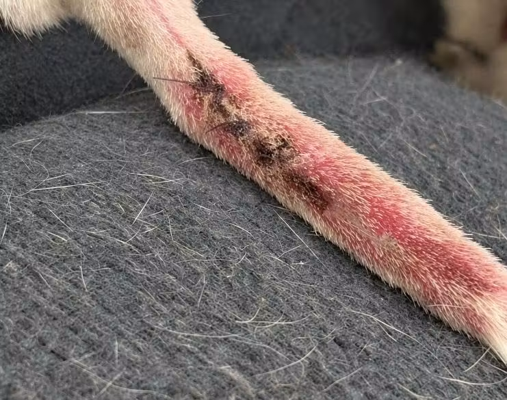
FEB 3 — Healing well
The tail was documented as healing well at this evaluation.
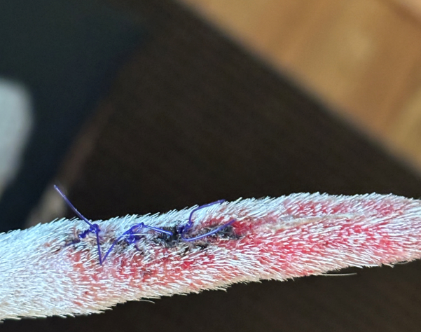
FEB 16 — Self-trauma noted
Phinney had self-traumatized the healing tail.
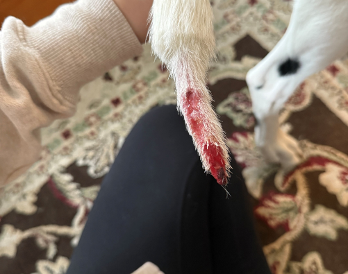
FEB 18 — Bone exposure; partial amputation recommended
Circulation to the tail tip was questionable, and bone was exposed. The area was clipped, cleaned, and bandaged, and we started antibiotics and pain medication.
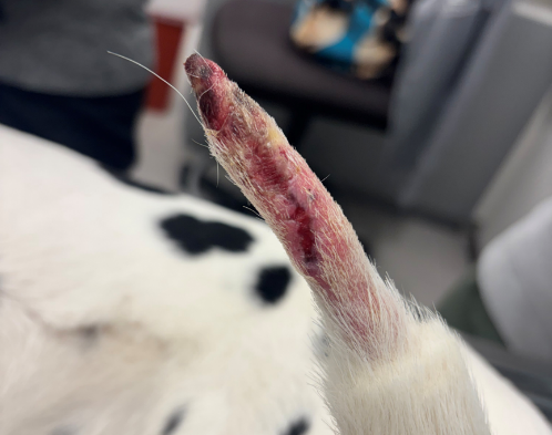
FEB 23 — MedcoVet Luma begins
Twice daily, 30 seconds per spot.
FEB 24 — Clinical laser treatment
FEB 25 — Amputation still recommended
Circulation to the tail tip remained questionable.
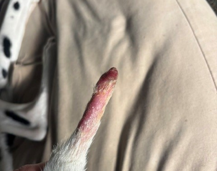
FEB 27 — Exudate and concern for infection
The tail remained inflamed and tender. Antibiotics continued, and amputation was still being considered.
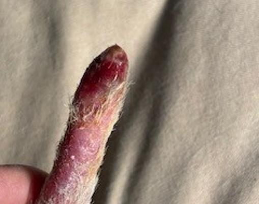
MAR 2 — Tail tip still at risk
The clinical team continued to monitor the wound and circulation.
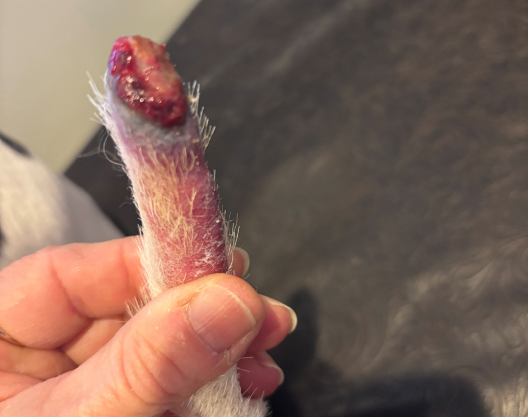
MAR 8 — Turning point: circulation improving
The wound was healing by second intention, circulation was improving, and the tail was less tender to the touch. Phinney could spend supervised time unbandaged. His veterinarian was hopeful but not yet certain the tail could be saved.
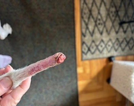
MAR 12 — Wound closing; no bone exposure
Phinney was almost back to normal activity. Manuka honey wraps (changed every 48 hours) and an Assisi Loop (three times daily) were added.
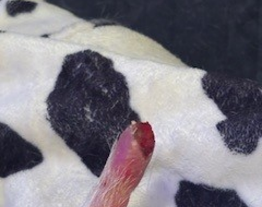
MAR 18 — Good skin coverage
The tail was smaller in diameter, although some swelling remained.
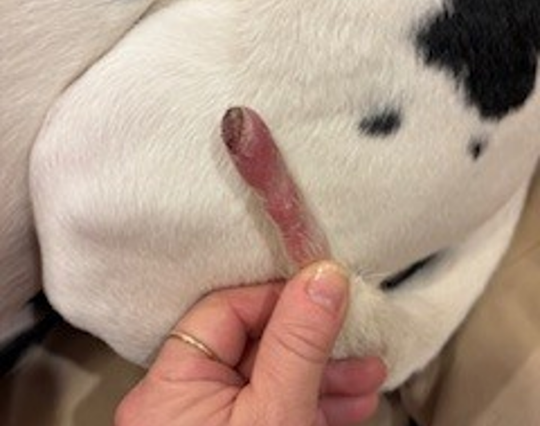
MAR 20 — Tail saved
Amputation was no longer necessary.
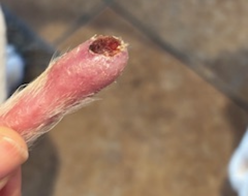
APR 27 — Completely healed
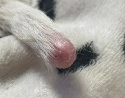
Outcome
Phinney kept his tail. On March 20, 25 days after MedcoVet Luma was added, amputation was no longer necessary, and on April 27, 2026, the tail was documented as completely healed.
The change was slow. Two weeks after Luma had begun, the tail tip was still in danger: amputation was advised on February 25, infection was a concern on February 27, and the loss of the tail tip was still possible on March 2. The point at which things began to turn around was on March 8, since circulation and tenderness were improving and the wound was healing by second intention. Further progress was made by the time the wound had closed on March 12 and by the time there was good skin coverage on March 18.
Since Phinney was given antibiotics, pain relief, bandaging, clinical laser treatment, Manuka honey, and an Assisi Loop together with MedcoVet Luma, this case is unable to assess the extent to which any one of the treatments made a contribution.
Case Status at Last Follow-Up
| Last documented follow-up | April 27, 2026 |
| Clinical status | Completely healed |
| Amputation | Not needed |
| Bone exposure | Resolved (none noted from March 12) |
| Activity | Almost back to normal by March 12; completely healed at the April 27 follow-up |
| MedcoVet Luma | Discontinued (April 27) |
Looking Back
The hardest part of this case was deciding whether to give the tail more time to heal or to go ahead with amputation. Since the wound was being closely watched and there was still a good chance of recovery, I thought it was worth trying a conservative approach. A dog’s tail is not just for expression and communication; it also helps with balance and movement. That’s why keeping as much of Phinney’s tail as possible mattered to me, as long as it was safe.
Consistent wound care, close monitoring, and adding Luma as a non-invasive treatment helped us support healing without another surgery for Phinney. Watching the wound heal completely and saving the tail made the decision very rewarding. Red light therapy can be a good option to help wound healing and should be considered as part of a full treatment plan when it is safe and appropriate.
“The tail is not merely cosmetic. It is an integral part of a dog’s communication and plays an important role in balance and coordinated movement. Seeing Phinney’s wound completely healed and knowing we were able to preserve his tail was incredibly rewarding.”
— Cathy Symons, CVT, CCRP, CHPT
Clinical Takeaways From Phinney’s Case
- A recommended amputation is not always the final outcome. Phinney’s tail had bone exposure and questionable circulation, yet with continued wound care and close monitoring, it healed completely.
- Early healing can be undone by self-trauma. The tail was healing well on February 3 before self-trauma on February 16 turned a simple wound into a complicated one. Preventing licking and chewing matters from the start.
- Improvement took time to show. Luma started on February 23, but amputation was still being discussed on March 2. Circulation and tenderness only clearly improved by March 8.
- Red light therapy fit into a multimodal plan. Luma was used alongside wound cleaning and bandaging, antibiotics, pain management, clinical laser, Manuka honey, and an Assisi Loop, not in place of them.
- Consistent home treatment made twice-daily sessions possible. Phinney’s owner, a veterinary technician, could deliver Luma at home twice a day without extra clinic visits.
- Serial photos and dated notes made the course clear. Twelve photos over three months let the clinical team track circulation, closure, and skin coverage and time the decision on amputation.
Phinney’s case does not establish a standard red light therapy protocol for tail wounds in dogs. It documents one dog with an exposed-bone tail wound, questionable circulation, and a recommended amputation, whose tail was saved and fully healed with a multimodal plan that included MedcoVet Luma.
Concurrent Treatment and Case Limitations
Phinney’s outcome needs to be read in the context of everything else he received.
MedcoVet Luma began on February 23, five days after antibiotics, pain medication, and bandaging started. A clinical laser treatment followed on February 24. Manuka honey wraps and an Assisi Loop were added on March 12, after the wound had already started to improve. Because these treatments overlapped, this case cannot determine how much of the improvement came from any one of them.
Other limitations consist of:
- This is a single patient case, not a controlled study.
- Healing was assessed from clinician and owner observation plus serial photographs, not standardized wound measurements or perfusion testing.
- It is not known whether Phinney’s tail would have healed with conventional wound care alone.
- Controlled studies of low-level laser therapy in healthy dogs have not shown a measurable effect on small, acute wounds (Kurach et al., 2015; Gammel et al., 2018).
- Response in other dogs may vary with wound location, depth, infection, circulation, self-trauma, and the other treatments used.
Any tail wound with heavy bleeding, swelling, or a change in tissue color needs prompt veterinary care (VCA Animal Hospitals). Red light therapy is an adjunct to veterinary wound management, not a replacement for it.
Frequently Asked Questions
Can a dog’s tail heal if the bone is exposed?
Sometimes. It depends on whether the tissue around the bone still has a blood supply and whether infection can be controlled. Phinney had exposed bone on February 18, and by March 12 no bone exposure was noted. A veterinarian needs to assess each case, because crushed bone or a wound that won’t heal may still need amputation (VCA Animal Hospitals).
How long does a dog tail wound take to heal?
It varies with the depth of the wound, circulation, infection, and whether the dog keeps reinjuring it. Phinney’s tail took about 12 weeks from the first injury to complete healing, including a setback from self-trauma. Wounds that keep getting hit by wagging may not heal until the trauma stops (VCA Animal Hospitals).
When does a dog need a partial tail amputation?
A vet might suggest a partial amputation of the tail in the case of a traumatic injury, if there has been repeated self-inflicted trauma, or when a wound fails to heal or keeps coming back even after conservative treatment (Risselada, Clinician’s Brief, 2022). Although it is a well-established procedure, it does require anesthesia and surgery.
Can red light therapy help a dog’s wound heal?
Red light therapy (photobiomodulation) is used as an adjunct in wound care because it increases cellular energy production and is able to reduce inflammation and oxidative stress (Hamblin, 2017). In dogs, two controlled studies found no measurable effect on small, acute wounds in healthy dogs (Kurach et al., 2015; Gammel et al., 2018), and complicated wounds have not been well studied. It is best used alongside, not instead of, standard wound management, as it was in Phinney’s case.
How do you stop a dog from reinjuring a tail wound?
Typical methods involve the use of a lightweight, self-adhesive bandage (one that is not tied on too tightly), an Elizabethan collar or a bitter spray to prevent the animal from chewing, and restricting access to small areas where the tail would hit against walls or furniture (VCA Animal Hospitals). The case of Phinney’s self-injury following early healing illustrates the importance of continued protection during recovery.
Is a dog tail injury an emergency?
Excessive bleeding, swelling, or a change in the color of the tail tissue are reasons to see a veterinarian right away (VCA Animal Hospitals). A tail that looks crushed or has exposed bone also needs prompt care.
Evidence Citations
Case study prepared by Cathy Symons, CVT, CCRP, CHPT, from her private practice. Published with pet parent consent.


