
CASE STUDY · FELINE DERMATOLOGY
Eosinophilic Granuloma Complex: How Red Light Therapy Helped One Cat Improve Without Long‑Term Steroids
SUMMARY
Trouble is a 5-year-old domestic shorthair cat who came to me with eosinophilic granuloma lesions in her mouth and throat, bad enough that eating and swallowing had become painful for her. She’d already been through a course of prednisolone that didn’t resolve them. Once a biopsy confirmed the diagnosis, I built her a MedcoVet Luma red light therapy protocol targeting both cheeks and lower jaw, with treatment directed toward the back of the mouth and throat. I started seeing improvement around weeks three to four. Trouble restarted prednisolone briefly mid-protocol for an allergy flare. Improvement became noticeable while prednisolone and Luma overlapped, so I can’t attribute that initial response to either treatment alone. What stood out clinically was that Trouble continued improving after prednisolone was discontinued while red light therapy continued. I have eight clinical photos documenting her healing from July 5 through August 7.
1
Biopsy-confirmed diagnosis
6 wks
Active protocol before taper
Wks 3–4
First visible improvement
8
Clinical photos, Jul 5–Aug 7
Case Snapshot
| Patient | Trouble, 5-year-old Domestic Shorthair cat |
| Condition | Eosinophilic Granuloma Complex: lesions, mouth and throat |
| Prior treatment | Prednisolone (lesions remained before Luma) |
| Protocol length | 6 weeks active treatment, ongoing maintenance |
| Outcome | Markedly improved, continued improvement after prednisolone was discontinued |
| Clinician | Kristy Williams, CVT, CCFT |
Baseline vs. Outcome
Trouble came to MedcoVet as a 5-year-old domestic shorthair with biopsy-confirmed feline eosinophilic granuloma complex. She had eosinophilic granuloma lesions affecting the back of her mouth and throat. Unlike the indolent ulcer, sometimes called a rodent ulcer, that commonly affects a cat’s upper lip, Trouble’s EGC lesions were located farther back in the oral cavity.
At baseline, the lesions were causing discomfort with eating and swallowing. Trouble also had a history of chronic allergy symptoms and overgrooming of her belly and the backs of her hind legs, with sores developing in those areas during flares. She had previously received prednisolone, but the lesions had not resolved with that treatment alone.
Before Treatment
- Biopsy-confirmed lesions in the cat’s mouth and throat
- Discomfort while eating
- Discomfort while swallowing
- Chronic allergic symptoms
- Overgrooming of the belly and backs of the hind legs, with sores during flares
- Previous prednisolone treatment without resolution of the lesions
- Goal: reduce the oral lesions and help Trouble return to comfortable, normal activity
After 6+ Weeks
- Lesions were markedly improved
- Serial clinical documentation showed gradual resolution of the oral lesions
- Improvement first became noticeable around weeks 3 to 4
- Prednisolone was completed around July 23
- Improvement continued after prednisolone was discontinued
- Luma treatment continued as part of Trouble’s long-term management
- Trouble’s clinician reported continued improvement beyond the initial six-week treatment period
Trouble’s case documents an uncommon use of red light therapy for a cat with eosinophilic granuloma complex, particularly because her lesions involved the mouth and throat rather than more familiar presentations such as an eosinophilic ulcer of the upper lip, eosinophilic plaque, or linear granuloma.
What Is Feline Eosinophilic Granuloma Complex? Clinical Signs Explained
Eosinophilic granuloma complex in cats is a group of immune-related skin and mucosal lesions, including the raised, yellowish-to-pink nodules called eosinophilic granulomas, that can show up in the mouth, throat, face, and other parts of the body, and make eating or swallowing painful (Merck Veterinary Manual; Cornell Feline Health Center).
It isn’t a single disease — it’s an umbrella term for three related skin and mucosal presentations in cats, grouped together because they share the same underlying immune mechanism.
Indolent (Rodent) Ulcer
Well-defined ulcerated area, often with raised borders and swelling, usually on the cat’s upper lip on one or both sides.
Eosinophilic Plaque
Raised, red, intensely itchy lesion. Most common on the belly and thighs.
Eosinophilic Granuloma
Raised, yellowish-to-pink nodule; can occur on the face, ears, paw pads, chin, tongue, mouth, hind legs, and thighs.
Trouble’s presentation
Trouble’s lesions fall into the granuloma category, affecting the mouth and throat rather than the more commonly discussed lip ulcers or oral ulcers. That’s part of why the presentation isn’t always immediately recognized by pet parents searching for “cat mouth sores,” “mouth ulcers,” or “rodent ulcer.”
What causes it: it is usually an allergic response and an overactive immune system — eosinophils release inflammatory chemicals in reaction to a perceived threat, and lesions can turn up at several common sites on the body, including the mouth and hind legs. Flea, mosquito, or mite bites, food allergy, dust mites, environmental allergens, and medication reactions can all trigger it, though in some affected cats I never land on a clear underlying cause. Identifying the trigger, when possible, helps guide treatment. In Trouble’s case, her pet parent reported that she had been on a hydrolyzed diet for many months without improvement, so an environmental allergy was suspected. Blood testing through Nextmune identified both food and environmental allergens, but no single trigger was considered definitively established.
Treatment depends on identifying triggers in affected cats, then controlling what you can. Flea control is crucial in treating eosinophilic granuloma complex, and a hypoallergenic diet trial makes sense if food allergy seems likely. Trouble had already completed a prolonged hydrolyzed diet trial without improvement. Oral immunotherapy was ordered after allergy testing but had not been started during the period documented in this case. For Trouble, she had already been down the standard medication path before I ever got involved.
The Challenge
Trouble is a young cat with a history of chronic allergies. During flares, she overgrooms her belly and the backs of her hind legs and develops sores in those areas. On top of that, she developed eosinophilic granuloma lesions in the back of her mouth and throat, which biopsy confirmed as eosinophilic granuloma complex, or EGC lesions.
- Lesions in the mouth and throat
- Discomfort while eating
- Discomfort while swallowing
- Chronic allergies
- Overgrooming and recurrent sores on the belly and hind legs
The main clinical signs here were pain with eating and swallowing.
Her pet parent’s goal was simple: clear the lesions so Trouble could get back to the normal activity expected of a young cat, without leaning on steroids indefinitely to manage a condition likely to recur with her allergies. I agreed that was the right goal — steroid-based care can quiet symptoms, but repeated use doesn’t address the underlying trigger, and it’s a poor long-term fit for a cat this young.
Previous Treatment With Prednisolone
Before she came to me, Trouble had already been treated with prednisolone — the standard first-line therapy for eosinophilic granuloma complex, and one that can resolve lesions for most cats; while many cats respond to it, Trouble’s oral inflammation and lesions endured before we started the red light therapy protocol.
Her pet parent recorded noticeable throat inflammation and swelling on June 10 after five days of prednisolone. Because of the seriousness and location of the lesions, a biopsy was planned to rule out cancer and confirm the EGC diagnosis before beginning red light therapy. Trouble also received a short-term steroid injection to help settle the inflammation quickly.
That history mattered again later in the case. Prednisolone was reintroduced mid-protocol during an allergy flare. At that point, the granulomas had not yet shown visible change with the initial once-daily Luma protocol. Untangling which treatment was doing what work is part of why I wanted to document this case carefully.
Trouble’s pet parent also crushed her hydrolyzed food into a pate to make eating easier while her mouth and throat were inflamed.
Why Avoiding Long-Term Steroids Mattered
Prednisolone works well for most cats, but once a cat needs more than three to four months of steroid therapy, VCA Animal Hospitals notes the condition should be re-evaluated or an alternative pursued, given a well-documented range of cumulative effects (VCA Animal Hospitals):
- Urinary tract infections, occurring in up to 30% of long-term patients
- Thinning skin, poor coat quality, and impaired wound healing
- Weight gain and muscle weakness from protein breakdown
- Increased susceptibility to bacterial and fungal infections
- Predisposition to diabetes mellitus and, in high-dose/long-term cases, medication-induced Cushing’s syndrome
(General veterinary literature on long-term corticosteroid use — not specific to Trouble’s case.)
For a 5-year-old cat with a lifetime of chronic allergy management potentially ahead of her, avoiding long-term steroid dependency was one reason her pet parent was looking for another option.
Diagnosis First
Before MedcoVet Luma treatment began, a biopsy was performed to confirm the diagnosis. This was a deliberate step, not a formality. Trouble’s throat had become inflamed and swollen, so the biopsy was used to rule out cancer as well as substantiate eosinophilic granuloma complex before red light therapy began. Skin biopsy and cytology are standard parts of the EGC diagnostic workup when a presentation isn’t classic (Merck Veterinary Manual).
EGC in this location wasn’t something I’d treated with red light therapy before. I was familiar with indolent (“rodent”) ulcers, the lip-ulcer subtype, but not with granulomas in the throat. So I researched the specific presentation and consulted MedcoVet’s laser therapy protocols before building Trouble a plan, rather than just applying a generic template.
Why I Chose Red Light Therapy for Trouble
Before contacting MedcoVet, Trouble’s specialist had discussed steroids and immunotherapy, with cyclosporine as a possible future option if her condition worsened. Her pet parent asked the specialist about red light therapy and shared a research article. The practice confirmed that in-clinic laser therapy was available, but repeated treatments would mean added cost, missed work, travel, and stress for Trouble. Her pet parent began researching at-home photobiomodulation therapy and found MedcoVet Luma.
I didn’t have much precedent to go on for this specific presentation. I’d used red light therapy successfully for indolent ulcers before, but not for granulomas in the mouth and throat. What I did know: photobiomodulation has a well-documented, low-risk safety profile, it’s non-invasive, and because the treatment areas were external — both cheeks and lower jaw, with treatment directed toward the back of the mouth and throat — it was something Trouble’s pet parent could actually deliver at home, consistently, without the stress or cost of driving Trouble in for repeated in-clinic sessions. The device (MedcoVet Luma) being 100% eye-safe mattered too, given how close we’d be working to her face.
Once the biopsy confirmed the diagnosis, I felt comfortable using red light therapy as a low-risk adjunct. I couldn’t know in advance whether Trouble would respond, which made close monitoring and regular communication with her pet parent especially important.
How Red Light Therapy Works
Red and near-infrared light therapy, also called photobiomodulation (PBM) or low-level laser therapy, works by delivering specific wavelengths of light that are absorbed by cytochrome c oxidase, an enzyme in the mitochondria of cells. That absorption increases ATP (cellular energy) production and triggers downstream effects including reduced oxidative stress, modulation of inflammatory signaling, and improved blood flow, mechanisms relevant to both wound healing and inflammation control (Previti et al., 2025). This is the mechanism I’m relying on any time I recommend MedcoVet Luma, whether that’s for arthritis, post-surgical healing, or, as with Trouble, an inflammatory mucosal condition like EGC.
Read more about the science of red light therapy here.
Treatment Protocol: How It Evolved
The protocol wasn’t set-and-forget. I adjusted duration and frequency based on how the eosinophilic granuloma lesions responded over time.
| Protocol Detail | Trouble’s Treatment |
|---|---|
| Treatment areas | Left and right cheek and lower jaw, with treatment directed toward the back of the mouth and throat. |
| Starting session time | Treatment began July 2 at 30 seconds per spot. |
| Starting frequency | Once daily. |
| First adjustment | After no visible change, treatment time was increased to 1 minute per spot, once daily before July 16. |
| Second adjustment | On July 16, treatment frequency was increased to twice daily for 5 days, while keeping treatment time at 1 minute per spot. |
| Why frequency increased | Trouble had shown no visible change with the initial once-daily protocol. The increase to twice-daily treatment was made to provide more frequent treatment to the affected areas. |
| Concurrent care | Trouble experienced an allergy flare and restarted prednisolone on July 13, three days before MedcoVet Luma frequency was increased. |
| When improvement began | The first significant improvement was seen during the period following the increase to twice-daily treatment. Because Trouble was also receiving prednisolone, I felt the improvement was likely related to both treatments. |
| Prednisolone discontinued | Trouble completed prednisolone around July 23. |
| Maintenance plan | After the active treatment period, Trouble transitioned to treatment every other day, using the same treatment areas at 1 minute per spot. She continued to improve after prednisolone was discontinued. |
How the Protocol Changed Over Time
| Stage | MedcoVet Luma Protocol | Clinical Response / Reason for Change |
|---|---|---|
| July 2 | 30 sec/spot, once daily | Initial protocol started |
| Before July 16 | 1 min/spot, once daily | Increased treatment time after no visible change |
| July 16 | 1 min/spot, twice daily for 5 days | Increased frequency because lesions had shown no visible change |
| Weeks 3-4 | Following twice-daily escalation | First significant improvement became noticeable; prednisolone was also being used during this period |
| Around July 23 | Prednisolone completed; red light therapy continued | Trouble continued to improve after medication ended |
| Maintenance | 1 min/spot, every other day, same treatment areas | Continued improvement and long-term management |
Current Maintenance Protocol
After the initial six-week treatment period, Trouble transitioned to an every-other-day maintenance schedule. On each treatment day, her MedcoVet Luma protocol includes treatment of both sides of the mouth:
Left Side
- Mouth: 2 spots, 1 min/spot, 2 treatments/day
Right Side
- Mouth: 2 spots, 1 min/spot, 2 treatments/day
The treatment areas remained consistent with the active protocol: the left and right cheek and lower jaw, with treatment directed toward the back of the throat.
Trouble’s maintenance plan is expected to be ongoing rather than a short-term course. Because eosinophilic granuloma complex can recur, her pet parent will continue to monitor her mouth closely, follow the maintenance protocol, and report any changes so treatment can be adjusted if needed.
Case Timeline & Photo Documentation
Eight clinical photos were submitted over five weeks, alongside the case’s key clinical events. A June 10 pre-Luma photo also documented severe throat inflammation after five days of prednisolone.
JUN 10 — Severe throat inflammation recorded
After five days of prednisolone. The degree of inflammation and swelling led to the decision to pursue biopsy to rule out cancer and confirm EGC before laser therapy.
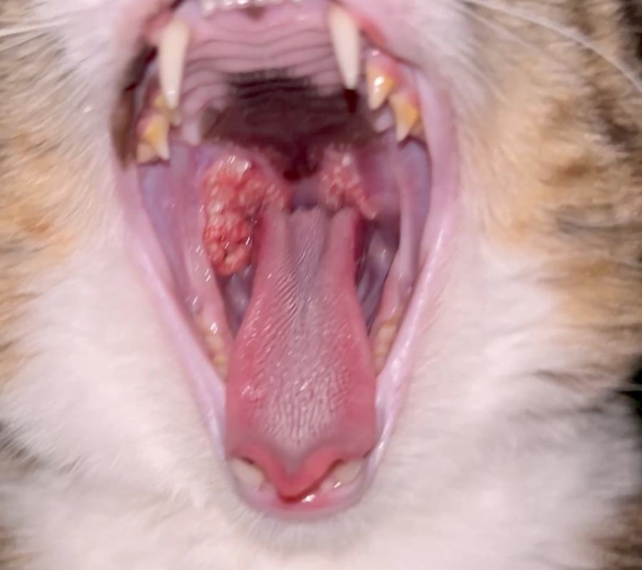
JUL 2 — MedcoVet Luma protocol begins
30 sec/spot, once daily, following biopsy-confirmed diagnosis
JUL 5 — Photo 1 submitted
Baseline documentation shortly after starting once-daily Luma sessions
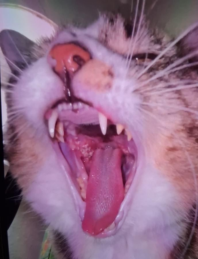
JUL 13 — Allergy flare; prednisolone restarted
Part of Trouble’s chronic allergy management during the treatment period
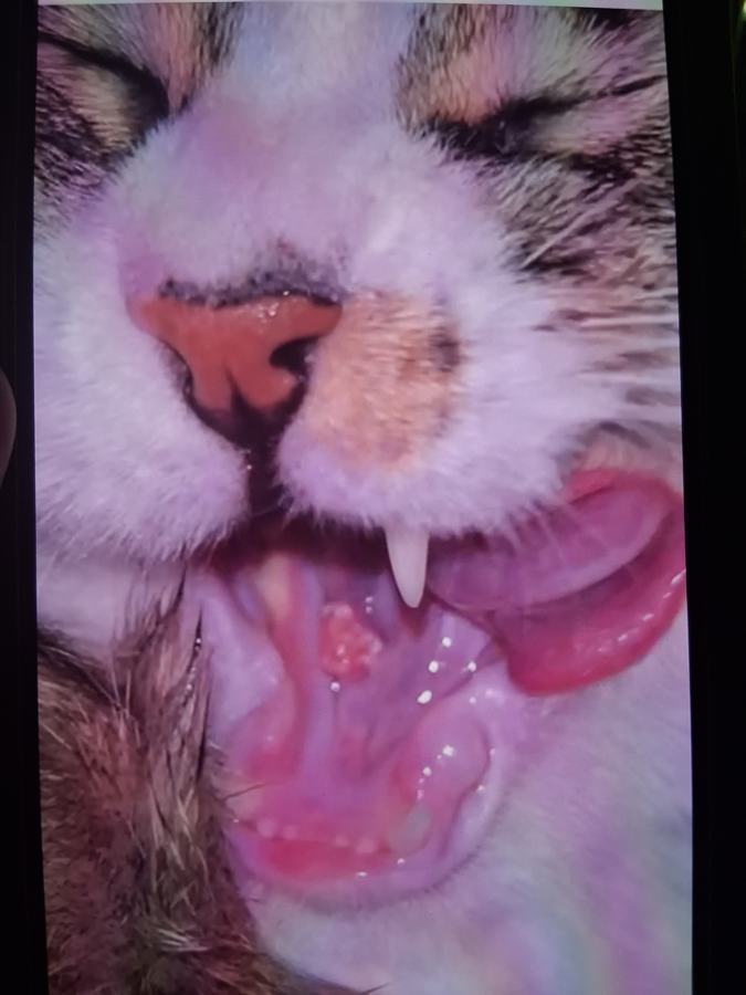
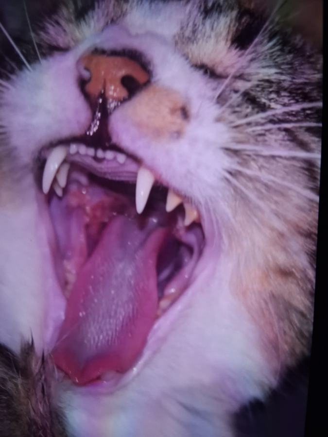
JUL 16 — Frequency increased to 2×/day
No change yet observed; escalated from once-daily for 5 days
JUL 18 — Photo 4 submitted
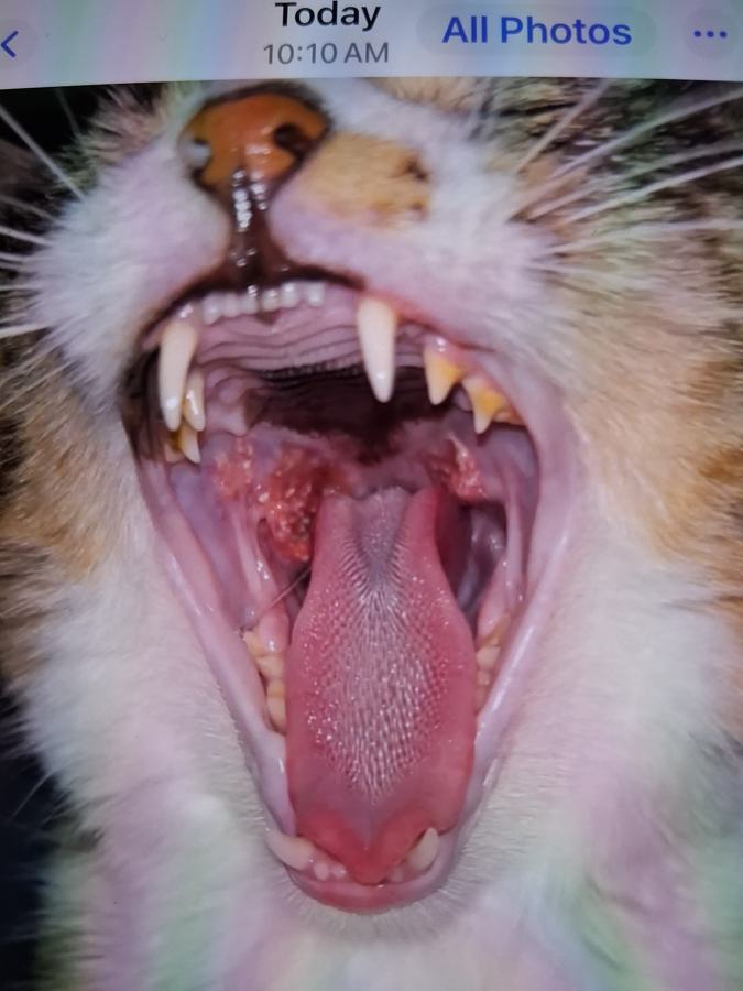
JUL 20 — Photo 5 submitted — first improvement window begins (weeks 3–4)
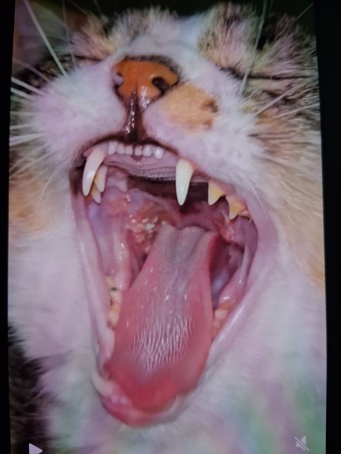
JUL 23 — Prednisolone discontinued
Every-other-day Luma maintenance begins
JUL 24 — Photo 6 submitted
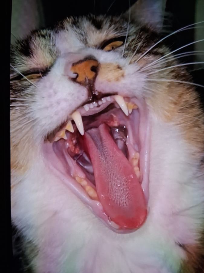
JUL 30 — Photo 7 submitted
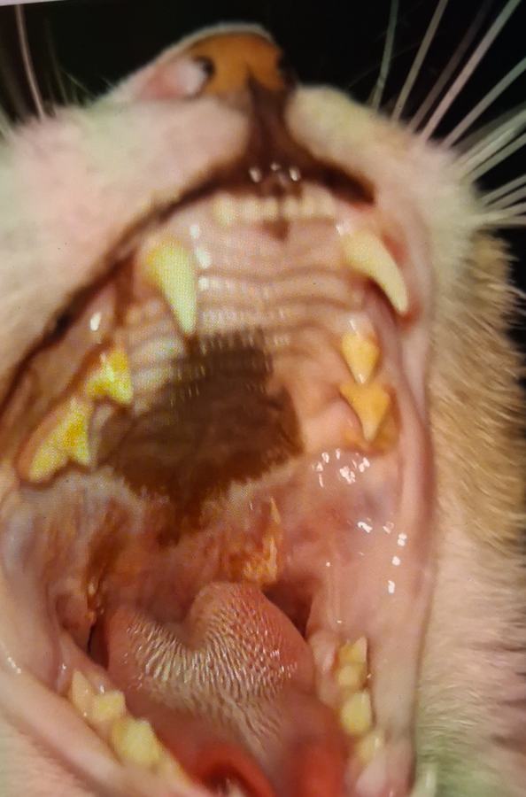
AUG 7 — Photo 8 submitted — continued improvement, most recent documentation
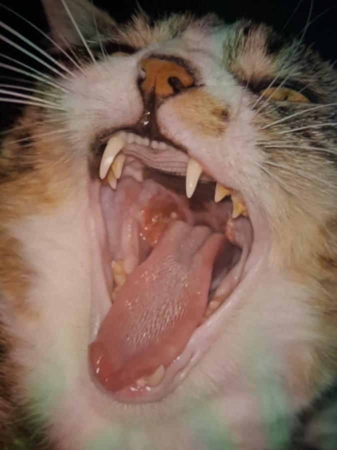
Outcome
By six weeks, Trouble’s lesions were markedly improved. She continued to improve after prednisolone was discontinued, while red light therapy remained part of her care plan.
That continued improvement was clinically encouraging, particularly because Trouble’s lesions remained even with the use of prednisolone before the MedcoVet Luma protocol. Because the two treatments overlapped during the period when improvement first became visible, this case cannot determine the individual contribution of prednisolone and red light therapy.
Trouble is now eating and swallowing comfortably and continues using MedcoVet Luma as part of her maintenance plan.
Trouble’s improvement does not mean her eosinophilic granuloma complex is permanently resolved. Her care is expected to require continued maintenance and close observation by her pet parent, with the goal of keeping the lesions controlled and responding early if signs begin to return.
Case Status at Last Follow-Up
| Last documented follow-up | August 7, 2026 |
| Clinical status | Markedly improved |
| Eating and swallowing | Comfortable |
| Prednisolone | Discontinued |
| MedcoVet Luma | Continuing as maintenance |
| Long-term goal | Maintain improvement, reduce recurrence |
Looking Back
This case worked out well, and I think that’s a big credit to Trouble’s owner. She was engaged and compliant from the start. She understood why the biopsy mattered before we began treatment, and she used a small towel during cheek treatments just in case Trouble moved, even though Luma is 100% eye-safe.
If I’m honest about what I’d change: looking back, I would have treated this condition as a wound from the start and gone straight to twice-daily sessions, rather than escalating from once daily.
Long-term, Trouble stays on an every-other-day maintenance schedule to help keep the lesions from returning.
Clinical Takeaways From Trouble’s Case
Trouble’s case provided several first-hand clinical observations that may help inform future use of photobiomodulation for cats with eosinophilic granuloma complex.
- Diagnosis came before treatment. Trouble’s diagnosis was confirmed by biopsy before Luma was started. Because oral ulcers, granulomas, swelling, and ulcerated areas in a cat’s mouth can have different causes, I wanted confirmation of the diagnosis before treating the tissue. This was particularly important given the location of Trouble’s lesions toward the caudal aspect of the mouth and throat.
- The initial once-daily protocol produced no visible change. Treatment began at 30 seconds per spot once daily and later increased to 1 minute per spot once daily. Trouble showed no noticeable change during the first two weeks.
- The protocol changed based on Trouble’s response. On July 16, Luma treatment increased to twice daily for five days because the lesions had not visibly changed. Treatment targeted the left and right cheek and lower jaw toward the back of the throat. This was the period when significant improvement first became apparent, although prednisolone had also been restarted three days earlier.
- I would start a similar case more frequently next time. Based on Trouble’s response, my main clinical takeaway was that I would approach a similar eosinophilic granuloma presentation more like a wound and begin twice-daily treatment earlier rather than starting once daily and increasing later.
- Concurrent prednisolone limits conclusions about cause and effect. Improvement became visible while both treatments were being used. I felt the response was likely related to both prednisolone and Luma. The case therefore supports documenting red light therapy as an adjunct used during treatment of feline eosinophilic granuloma complex, rather than claiming that it independently resolved the lesions.
- Improvement continued after prednisolone ended. Prednisolone was completed around July 23. Trouble continued red light therapy and continued improving after the medication was stopped. That ongoing response supported continued use of Luma as part of her maintenance plan.
- Consistent home treatment was a major part of the case. Trouble’s pet parent was highly engaged and followed the protocol closely. That consistency allowed me to assess changes over time and adjust frequency when the initial protocol produced little visible response.
Trouble’s case does not establish a standard red light therapy protocol for eosinophilic granuloma complex in cats, but it provides a documented clinical use case involving a young cat with biopsy-confirmed oral EGC lesions, previous steroid use, protocol adjustments, concurrent treatment, and continued improvement beyond six weeks. For future cases, I would begin with a more frequent treatment schedule while continuing to account for the underlying allergy, lesion location, other treatments, and the individual cat’s response.
Concurrent Treatment and Case Limitations
Trouble’s improvement needs to be considered in the context of the other treatment she received during the case.
Luma treatment began on July 2 at 30 seconds per spot once daily. When no visible change was seen, treatment increased to 1 minute per spot once daily. Trouble then experienced an allergic flare and restarted prednisolone on July 13. On July 16, because the EGC lesions had still shown little visible change, Luma frequency was increased to twice daily for five days.
This was also the period when Trouble began showing more significant improvement.
Because prednisolone and red light therapy overlapped, this case cannot determine how much of the initial improvement came from either treatment individually. My clinical assessment is that both likely contributed. This is especially important when interpreting a single case involving an inflammatory condition such as eosinophilic granuloma complex, where changes in the underlying allergic response, medication use, and other factors may all affect the lesions.
Prednisolone was discontinued around July 23. Trouble continued receiving red light therapy and continued to improve afterward. That continued improvement made ongoing treatment clinically interesting, particularly because her lesions had persisted despite prednisolone before the Luma protocol. However, a single case does not establish red light therapy as a standalone treatment for granuloma complex in cats, nor does it show that red light therapy will allow other affected cats to discontinue oral medications.
Other limitations include:
- This is a single patient case, not a controlled study.
- Trouble received concurrent medication during part of the improvement period.
- The outcome was based largely on clinician and pet-parent observation plus serial clinical images, rather than standardized lesion measurements.
- Trouble has chronic allergies. A prolonged hydrolyzed diet trial did not improve her signs, and an environmental allergy was suspected. Blood testing identified both food and environmental allergens, but no single underlying trigger was considered decidedly proven.
- Oral immunotherapy had been ordered but was not started during the period documented in this case, so it did not contribute to the improvement shown here.
- Response in other cats may vary based on lesion type, location, severity, allergy triggers, external parasites, food allergy, other underlying conditions, and the treatment used alongside photobiomodulation.
For cats with oral lesions, proper diagnosis remains important. Mouth ulcers, swelling, ulceration, or other changes in a cat’s mouth can have many causes, including dental disease, inflammatory disease, infection, trauma, and conditions such as squamous cell carcinoma. Trouble’s diagnosis was confirmed by biopsy before red light therapy began so the clinical team knew what they were treating.
Frequently Asked Questions
Is eosinophilic granuloma complex the same as a rodent ulcer in cats?
Not exactly. A rodent (indolent) ulcer is one of three lesion types within eosinophilic granuloma complex, typically appearing on the upper lip. EGC also includes eosinophilic plaques and granulomas, which can affect the belly, thighs, mouth, and throat, as in this case.
What does eosinophilic granuloma look like in cats?
Presentation depends on the subtype: indolent ulcers are well-defined red sores usually on the upper lip; plaques are raised, intensely itchy red lesions on the belly or thighs; granulomas are raised yellowish-to-pink nodules that can appear on the face, ears, paw pads, chin, mouth, or thighs, with some oral presentations also showing ulceration.
How long does it take to see improvement with red light therapy for EGC in cats?
In this case, visible improvement appeared around weeks 3–4 of daily-to-twice-daily treatment, with the most notable phase following the escalation to twice-daily sessions. Timelines will vary by severity, lesion type, and treatment consistency.
Can eosinophilic granuloma in cats be treated without long-term steroids?
Treatment depends on the underlying cause and severity, though corticosteroids remain the standard first-line treatment and work for most cats. This case suggests red light therapy may help some cats reduce dependence on steroids, but that hasn’t been established in controlled studies for EGC specifically, and severe cases may need more aggressive management. Talk to a clinician about what’s appropriate for your cat.
Is eosinophilic granuloma complex in cats contagious or dangerous?
It isn’t contagious to other pets or people. Progression to cancer is extremely rare, even for indolent ulcers, which historically raised that concern. Left untreated, lesions can cause ongoing discomfort and, in mouth/throat cases like Trouble’s, interfere with eating. More broadly, the most common cause of mouth ulcers is dental disease rather than EGC. Routine dental care may help prevent mouth ulcers in general, and dental diets can be part of overall oral-health support.
Evidence Citations
Case study prepared from clinician-submitted case documentation (Case #5911), MedcoVet. Published with pet parent consent.


