
The WAIT Test™ › Irradiance
Irradiance in Red Light Therapy: What It Means, Why It Matters, and How to Evaluate a Device
On this page
- Why is irradiance important?
- What does irradiance mean in the context of red light therapy?
- Where Irradiance fits in The WAIT Test™
- How to evaluate an irradiance claim
- What changes when the patient has fur?
- The WAIT Test™ standard for Irradiance
- Strong disclosure, incomplete disclosure, and warning signs
- Worked example: usable light at the hip
- The science: how irradiance sets the dose
- Irradiance explained: the turkey analogy
- Irradiance units and conversions
- How irradiance connects power, area, time, and radiant exposure
- Irradiance is not average power
- Irradiance is not the dose
- Equal J/cm² does not mean an equal biological effect
- Why irradiance matters biologically
- Two irradiance ranges, two different jobs
- Is there an ideal irradiance for red light therapy?
- What veterinary research tells us about irradiance
- Technical details
- Thermal effects: PBM is not defined by a temperature reading alone
- Does higher irradiance send light deeper?
- Why distance changes irradiance
- Peak, average, and spatial irradiance are different
- Spatial uniformity: one number can hide the treatment area
- Beam area must be defined at the treatment plane
- Multi-wavelength devices need more than one combined figure
- Irradiance is an energy rate, not a photon count
- Laser and LED irradiance cannot be compared by one number alone
- How irradiance should be measured
- Why lux, lumens, and phone apps cannot replace irradiance
- Measured output matters more than component ratings
- Frequently asked questions about irradiance
- What is a good irradiance for red light therapy?
- Is 100 mW/cm² too high?
- Is 10 mW/cm² too low?
- Can high irradiance relieve pain?
- Is irradiance the same as power density?
- Is irradiance the same as energy density?
- Can I calculate irradiance from watts?
- Can I figure out treatment time using irradiance?
- Will a higher irradiance result in a shorter treatment?
- Does higher irradiance go further?
- What causes irradiance to decrease as the distance increases?
- Is the center reading the irradiance?
- How is average irradiance different from peak irradiance?
- Can a lux meter be used to measure the irradiance of red light therapy?
- Can two devices that have the same irradiance provide different treatments?
- Does pulsing make high peak irradiance more effective?
- What amount of irradiance reaches a joint?
- What is the relevance of fur when the open-air irradiance is high?
- Should irradiance be measured through a spacer or cover?
- The bottom line
- Continue The WAIT Test™
- Research and references
Irradiance is the concentration of light or the amount of light power that lands on each square centimeter of the treatment surface, measured in milliwatts per square centimeter (mW/cm²). It’s the number you need to work out treatment time and dose.
Why is irradiance important?
Because irradiance and time together make the dose. The dose, measured in joules per square centimeter (J/cm²), is simply irradiance multiplied by treatment time:
Dose (J/cm²) = irradiance (W/cm²) × time (seconds)
For example, 50 mW/cm² for 80 seconds delivers 4 J/cm². So does 100 mW/cm² for 40 seconds. Without accurate irradiance, you can’t know the dose your pet receives or set the right treatment time.
But PBM doesn’t reliably follow reciprocity: you can’t trade irradiance for time and expect the same result. The same dose delivered at different irradiances produces different effects. Too little irradiance, and cells barely respond no matter how long you wait. Too much, and the benefit can level off or even reverse (Huang et al., 2011). Around 50 to 100 mW/cm², light stimulates cells and calms inflammation. At much higher irradiance, often hundreds of mW/cm² at the nerve, it can temporarily slow pain signals. Irradiance also affects how quickly heat builds up.
What does irradiance mean in the context of red light therapy?
The quick answer
Irradiance is the amount of radiant power reaching a surface divided by the area of the illuminated surface. It is generally referred to as power density and is normally given in milliwatts per square centimeter (mW/cm²).
Irradiance (W/cm²) = incident optical power (W) / illuminated area (cm²)
When 1 W is distributed evenly over 10 cm², the average irradiance is 0.1 W/cm², or 100 mW/cm²; if the same 1 W is spread evenly over 100 cm², the average irradiance is 0.01 W/cm², or 10 mW/cm².
The amount of power is the same. The concentration per square centimeter is ten times higher in the first case.
When evaluating a product, look for information in two parts.
Part 1: The basics you can check yourself. These three numbers should be listed on the product page or in the manual:
- Stated irradiance in mW/cm²
- Spot size (treatment area) in cm²
- Average power in W or mW
With these, you can do two quick checks. First, make sure they agree: irradiance should be roughly the average power divided by the spot size. For example, 1 W (1,000 mW) over 14.5 cm² is about 69 mW/cm². Second, see where the irradiance falls: in the stimulation range of about 50 to 100 mW/cm², or much higher (hundreds of mW/cm²) for short-term pain relief (both described in “Two irradiance ranges, two different jobs” below).
For most devices, these basics are enough to screen out obvious problems. If they’re missing or don’t add up, that’s already your answer.
Part 2: The complete picture. These details are more technical, and you may need to ask the company for them:
- Where the irradiance was measured: in contact with the skin, or at a stated distance
- Treatment time and the resulting dose in J/cm²
- Which wavelengths are on, and the irradiance of each if there’s more than one
- Continuous or pulsed output, and confirmation that the irradiance is an average, not a peak
- Whether the figure is an average across the treatment area, or a center, peak, or minimum reading
- An irradiance map or other evidence that the light is spread evenly
- The measurement instrument, method, calibration, and tolerance
- Whether output stays steady through a full session
- Whether covers, lenses, or spacers were in place during measurement
A strong company can answer both parts. An irradiance figure without the Part 1 basics won’t give you a usable picture at all.
Where Irradiance fits in The WAIT Test™
Red light therapy has one job: get the right kind of light, in the right amount, to the target tissue. That’s why MedcoVet built The WAIT Test™.
The WAIT Test™ checks whether a red light therapy device can deliver a useful dose of light to the tissue that needs it. It has four components, and a device must pass all four:
Wavelengths
Is it the right kind of light for the target tissue?
The device must emit red or near-infrared light in a band that counts for the condition.Read the guide →Average Power
Is there enough light to treat the whole area?
The device’s true average optical output must cover the treatment area in one session.Read the guide →Irradiance
Is the light concentrated enough to deliver the dose in time?
The usable light at the skin must deliver the dose at each site within the time limit.You’re reading this guideTarget Tissue
Does the light reach the target tissue?
The light must get past fur and be applied the way the condition needs.Read the guide →Each device is judged separately for each condition and coat, so the same device can pass for a wound and fail for a hip. The full method, including the data you need and the decision rules, is in The WAIT Test™ guide.
This guide covers the third component, Irradiance. Irradiance turns a device’s power into a dose at the skin, but it only helps if the light is the right kind, the power behind it is honest, and the light actually reaches the target tissue.
The central question is:
Does the device give a measured irradiance that represents the area at the actual treatment surface, and does that measurement support a clear treatment time and a reasonable protocol for the tissue being treated?
This guide starts with how to check a device’s irradiance, including a worked example. Then it explains how irradiance and time set the dose, and it ends with technical detail on distance, uniformity, and measurement.
How to evaluate an irradiance claim
Keep it simple. Three steps, plus an optional fourth:
Step 1: Find the three key numbers
Look on the product page or in the manual for:
- Irradiance in mW/cm²
- Spot size (treatment area) in cm²
- Average power in W or mW
If any of these are missing, that’s already a warning sign.
Step 2: Check that they add up
Irradiance should be roughly the average power divided by the spot size. For example, 1 W (1,000 mW) over 14.5 cm² is about 69 mW/cm². If the numbers don’t agree, the company may be quoting peak power, electrical wattage, or a reading from a single bright spot.
Step 3: See which range the irradiance falls in
Is it in the stimulation range of about 50 to 100 mW/cm², or much higher (hundreds of mW/cm²) for short-term pain relief? Make sure that matches the job you want the device to do.
Then allow for the coat. If the device sits on top of the fur, The WAIT Test™ counts only a quarter of its irradiance at the skin. A device listed at 60 mW/cm² delivers about 15 mW/cm² of usable light through a light coat, and only about 5 mW/cm² through a dark one.
Optional step 4: Ask for a third-party test report
For extra confidence, you can ask the company for an independent, third-party report that measures the device’s output. A useful report shows the measured average power, the irradiance at the skin or treatment distance, and the spot size, along with the instrument and method used. It’s a nice extra, not a requirement. Plenty of good devices are made without one.
Then complete the rest of The WAIT Test™. Irradiance can’t fix the wrong wavelength, an inaccurate average power, or light blocked by fur.
What changes when the patient has fur?
An open-air irradiance measurement describes what arrives at the detector under those conditions. It doesn’t show what reaches a pet’s skin through a coat.
The WAIT Test™ handles this with a simple rule: usable light at the skin = device irradiance × coat color factor × fur factor. If the device doesn’t part the fur, only 25% of the light gets through the coat. A dark (brown) coat passes 33% of the light a light coat does; black coats pass less still and aren’t scored separately. Wounds are clipped, so none of these losses apply. For a deep target, the device counts only the near-infrared share. The coat-color effect is based on the same 47-dog study (Hochman-Elam et al., 2020).
This is why you must read irradiance with the final part of The WAIT Test™: Target Tissue.
The standard
The WAIT Test™ standard for Irradiance
The Irradiance component has four checks, worth 35 points in all. Each is scored separately for every condition and coat.
- I1. Irradiance known or calculable (10 points). The irradiance is known from the company, a MedcoVet measurement, or a third party, or it can be calculated from average power and head area.
- I2. Measurement conditions stated (5 points). The source says where the irradiance was measured: the distance and the area.
- I3. Delivers the dose per site in time (10 points). Using the usable light at the skin, the device must deliver WALT’s target dose at each treatment site within 5 minutes (30 for wounds).
- I4. Enough light at the skin, within the safe range (10 points). The usable light at the skin must be enough for 5 mW/cm² to reach the target’s depth: 5 mW/cm² for wounds, about 11 for most joints and the spine, 17 for the shoulder, and 25 for the hip. The device must also stay under the test’s irradiance cap unless the company gives thermal guidance.
Company specifications, MedcoVet measurements, and third-party sources all count. When the red/near-infrared split or head area isn’t known, usable light and time per site are worked out at best and worst case.
Irradiance result
Pass or fail, per condition. I1, I3, and I4 are worth 10 points each, so failing any one of them fails Irradiance. I2 is worth 5 points: missing it costs points but still passes.
How The WAIT Test™ scores a device. Each device gets one result for each situation (the condition being treated and the coat over the area), not one result overall. Each check is worth a set number of points, and the checks are grouped under the four components. A device passes only if it clears the eye-safety gate and earns at least 75% of the available points in each of W, A, I, and T. It fails if any component stays below 75% even when every missing figure turns out as favorably as possible. If nothing has failed but a missing figure could still decide the result, it needs more information: it can’t pass until the figure is supplied, but it hasn’t failed either. The score out of 100 ranks devices and never overrides the result; when figures are missing, it’s shown as a range. Class 3B and 4 lasers and Risk Group 2–3 LEDs are not safe for home use and aren’t scored. See how every device does, condition by condition, in The WAIT Test™ Device Scorecard.
If the information isn’t available
If irradiance isn’t known and can’t be calculated, I1, I3, and I4 are open. The device needs more information unless another component has already failed.
If irradiance is known but the red/near-infrared split isn’t, usable light for a deep target runs from zero to the full figure. A device whose best case still falls short fails. One whose best case clears the bar needs more information until the split is published.
The one question to ask
What are the irradiance, spot size, and average power of this device?
If the irradiance isn’t known and can’t be calculated, I1 stays open, and the device can’t pass the Irradiance part of The WAIT Test™ until it is.
Strong disclosure, incomplete disclosure, and warning signs
Strong irradiance disclosure
Irradiance: 76 mW/cm² at the skin. Spot size: 14.5 cm². Average power: 1.1 W. Measured by an independent lab; report available on request.
All three numbers are listed; they add up (1.1 W ÷ 14.5 cm² ≈ 76 mW/cm²), and the irradiance sits in the stimulation range. The third-party report is a bonus.
Incomplete disclosure
Irradiance: 100 mW/cm².
There’s no spot size or average power to check it against, and no report to confirm it.
Warning signs
Be cautious when a company:
- Doesn’t list irradiance, spot size, or average power
- Lists numbers that don’t add up
- Quotes peak power or electrical wattage instead of average power
- Lists an irradiance far outside the range for the job it claims to do
- Claims that more mW/cm² always means deeper or better treatment
Worked example
Worked example: usable light at the hip
Here’s how the Irradiance checks play out for four devices in The WAIT Test™ device comparison, for a 50 lb dog’s hip under a light coat. The hip needs 9 J/cm² at each site within 5 minutes, and at least 25 mW/cm² of usable light at the skin.
| Device | Irradiance at the device | Usable light at the skin | Time per site (I3) | Enough at the skin? (I4) | Irradiance |
|---|---|---|---|---|---|
| MedcoVet Luma | 89.7 mW/cm² | 89.7 mW/cm² (parts the fur) | 1.7 min | Yes | 100%: pass |
| Novaa Light Pad | 120 mW/cm² | 0–30 mW/cm² (on the fur; split not published) | At least 5 min | Depends on the split | 43–100%: need more info |
| Hooga Red Light Therapy Mat | 60 mW/cm² | 0–15 mW/cm² (on the fur; split not published) | At least 10 min | No | 43%: fail |
| B-Cure Laser Vet Pro | 20.2 mW/cm² | 5.1 mW/cm² (on the fur) | 29.6 min | No | 29–43%: fail |
The Novaa and Hooga pads both rest on the coat, and neither publishes its red/near-infrared split, so their usable light runs from zero (no near-infrared) to a quarter of the rated figure (all near-infrared). Even in the best case, the Hooga needs 10 minutes per site and falls short of 25 mW/cm², so it fails. The Novaa’s best case just reaches the limits, so it needs more information: publishing its near-infrared share would settle it. The Luma delivers about three times the Novaa’s best case.
The same rules apply to our own device. Under a dark coat, the Luma’s usable light drops to about 29.6 mW/cm², and each hip site takes 5.1 minutes, just over the 5-minute limit. It fails I3, and so fails Irradiance in that situation.
Deep dive
The science: how irradiance sets the dose
Irradiance isn’t a simple concept. It’s easy to confuse with power, dose, or brightness, and the same number can mean very different things depending on how it was measured. Fortunately, there’s a simple way to understand it.
Irradiance explained: the turkey analogy
One of the easiest ways to understand why irradiance matters is the turkey analogy.
Roasting a turkey depends on the same two things as red light therapy: energy intensity and how long you apply it. In the kitchen, that’s oven temperature and cooking time. In red light therapy, it’s irradiance and treatment time. And just like in the kitchen, getting the total right isn’t enough. You need the right intensity for the right amount of time.
Here’s how it plays out.
To roast a turkey, you usually set the oven to about 400°F and cook it for about 3 hours. Simple.
So why not crank the oven to 10,000°F and be done in about 7 minutes? Or set it to 1°F and wait 1,200 hours, which is 50 days? On paper, all three deliver the same amount of cooking: temperature × time = 1,200 “degree-hours.” In real life, the 10,000°F turkey chars to ash on the outside and stays raw in the middle. The 1°F turkey is still raw seven weeks later. Only the 400°F oven gives you dinner.
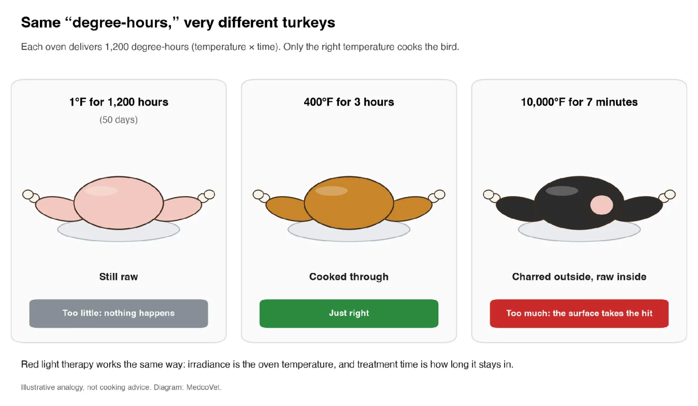
Figure 1. Three ovens, the same 1,200 degree-hours, three very different results. Only the right temperature, for the right time, cooks the bird. Diagram: MedcoVet.
That’s irradiance. Oven temperature is irradiance: the right amount of heat, or, in red light therapy, the right amount of light hitting each square centimeter. The time is how long you apply it. Temperature and time together make the dose. Get the temperature wrong, and no amount of time will fix it.
Here’s the same 1,200 degree-hours at five different oven settings:
| Oven temperature | Time for 1,200 degree-hours | What you get | The red light therapy version |
|---|---|---|---|
| 1°F | 1,200 hours (50 days) | Frozen and raw | Far too little irradiance: no useful effect, and sessions no one can stick with |
| 100°F | 12 hours | Warm, raw, and unsafe to eat | Too little irradiance: very long sessions for little benefit |
| 400°F | 3 hours | Golden and cooked through | The right irradiance for the right time |
| 1,000°F | 72 minutes | Burnt outside, undercooked inside | Too much irradiance: the surface takes the hit, heat builds, and the benefit can drop off |
| 10,000°F | About 7 minutes | Ash outside, raw inside | Far too much: all surface, no benefit |
If you read our Average Power post (part of the WAIT Test)
Here’s where power fits. Power is the oven’s heating element. A bigger oven needs a bigger, hotter element to reach the same temperature.
Nobody asks how hot the heating element gets. But if that number is wrong, you can’t work out the oven temperature. Average power is the same: you rarely think about it, but it’s the input you need to calculate irradiance.
| In the kitchen | In red light therapy |
|---|---|
| Heating element | Average power (W) |
| Size of the oven | Treatment area (cm²) |
| Oven temperature | Irradiance (mW/cm²) |
| Cooking time | Treatment time (seconds) |
| Temperature × time | Dose, or radiant exposure (J/cm²) |
| How done the turkey is | The biological response |
| Bake vs. broil | The two irradiance ranges (see “Two irradiance ranges, two different jobs”) |
Cells respond to light much the way a dish responds to heat. They need the right amount of light, at the right rate, for the right amount of time. Too little, and nothing happens. Too much, and the effect can level off or even reverse (Huang et al., 2011).
Irradiance units and conversions
The most common units are W/cm² and mW/cm².
- 1 W = 1,000 mW
- 1 W/cm² = 1,000 mW/cm²
- 100 mW/cm² = 0.100 W/cm²
- 75 mW/cm² = 0.075 W/cm²
- 10 mW/cm² = 0.010 W/cm²
Getting the units wrong can throw off your calculation by a factor of 1,000. Always convert milliwatts to watts before multiplying by seconds to get J/cm².
Some technical sources use W/m². Since 1 m² contains 10,000 cm²:
- 1 W/cm² = 10,000 W/m²
- 1 mW/cm² = 10 W/m²
Make sure you use the same area unit throughout your calculation.
People often use the word ‘intensity’ to mean brightness, power, irradiance, or even a device setting. This term is too vague for technical details. Always ask for the exact measurement and its unit.
‘Energy density’ usually means J/cm², but the correct term is radiant exposure. ‘Power density’ is another name for irradiance. Using the right terms helps avoid confusing watts and joules, which is more than just a wording mistake.
This problem is common in published literature and product marketing. A systematic review of 74 PBM papers found that 43% did not report irradiance, 40% did not report beam area, and 73% did not explain how they measured the light. The authors concluded that missing and inaccurate parameters impair repeatability and reliability (Hadis et al., 2016).
How irradiance connects power, area, time, and radiant exposure
For a uniform beam or for a clearly defined area average:
Average irradiance = average optical power / illuminated area
Treatment time converts irradiance into radiant exposure:
Radiant exposure (J/cm²) = average irradiance (W/cm²) x time (s)
Rearranging the equation gives the time required for a selected surface exposure:
Time (s) = radiant exposure (J/cm²) / irradiance (W/cm²)
A worked example
Suppose a contact applicator has:
- Measured average optical power: 1.10 W
- Treatment area at the skin: 14.5 cm²
- Treatment time: 60 seconds
If the power is distributed evenly across that area:
1.10 W / 14.5 cm² = 0.0759 W/cm² = 75.9 mW/cm²
The surface radiant exposure after 60 seconds is:
0.0759 W/cm² x 60 s = 4.55 J/cm²
The total radiant energy is:
1.10 W x 60 s = 66 J
The two paths agree: 66 J divided by 14.5 cm² equals about 4.55 J/cm².
However, this calculation assumes the measured power reaches the area you measured, the area is in the same plane, and the output is fairly even. In reality, a beam might have a bright center, weaker edges, gaps, or several hot spots. The math gives you an average, not the exact exposure at every spot.
Irradiance is not average power
Average power is the total rate of optical energy output. Irradiance is the rate per unit area at a specified plane.
Two products can have the same power and very different irradiance:
| Average power | Treatment area | Average irradiance |
|---|---|---|
| 1 W | 5 cm² | 200 mW/cm² |
| 1 W | 20 cm² | 50 mW/cm² |
| 1 W | 100 cm² | 10 mW/cm² |
The reverse is also true. Products can have the same irradiance and very different total power because they cover different areas:
| Average irradiance | Treatment area | Total power |
|---|---|---|
| 50 mW/cm² | 5 cm² | 0.25 W |
| 50 mW/cm² | 20 cm² | 1.00 W |
| 50 mW/cm² | 100 cm² | 5.00 W |
That’s why you shouldn’t compare wattage or irradiance without knowing the area. A focused probe and a large panel serve different coverage needs.
Irradiance is not the dose
Irradiance is a rate. Radiant exposure is the time-integrated surface exposure.
- Irradiance: J/s/cm², which is W/cm²
- Radiant exposure: J/cm²
If someone says a device gives a ‘dose of 75 mW/cm²,’ they’re mixing up a rate with a total amount. If they say it provides ‘8 J/cm²’ but don’t mention treatment time, you don’t know how fast the energy is delivered.
A complete surface-exposure statement needs both:
75 mW/cm² for 60 seconds = 4.5 J/cm²
It must also include the wavelength, power, irradiation time, beam area, pulse mode, treatment frequency, and anatomical location. Recommendations on reporting these closely related parameters have emphasized them for over ten years (Jenkins and Carroll, 2011).
Equal J/cm² does not mean an equal biological effect
In elementary radiometry, doubling irradiance and halving time preserves radiant exposure:
| Irradiance | Time | Radiant exposure |
|---|---|---|
| 40 mW/cm² | 100 s | 4 J/cm² |
| 100 mW/cm² | 40 s | 4 J/cm² |
| 200 mW/cm² | 20 s | 4 J/cm² |
The joules per square centimeter match. The rate and duration do not. It’s the turkey again: the same degree-hours don’t guarantee the same dinner.
Biological reciprocity means that two exposures with equal irradiance multiplied by time produce the same biological outcome. PBM does not follow that rule, except perhaps within a narrow range of closely matched settings. In a mouse pressure-ulcer model, researchers held radiant exposure at 5 J/cm² per day while changing irradiance, exposure time, and treatment schedule. The healing outcomes differed in this model (Lanzafame et al., 2007). Cultured human gingival fibroblasts have also shown different proliferation responses when equal radiant exposure was delivered at different irradiances (Almeida-Lopes et al., 2001).
Possible reasons include:
- Cellular signaling may depend on both rate and duration
- Photochemical intermediates may be created and cleared during exposure
- Oxygen availability can change during irradiation
- Heating can differ even when total J/cm² matches
- Pulsing can change the timing of light delivery
- Very short exposures may not engage the same sequence of events as longer ones
The key takeaway is simple but important: don’t assume any mix of irradiance and time will work just because the J/cm² total matches. A good protocol should clearly state both values.
Why irradiance matters biologically
PBM often shows a biphasic response: too little exposure may produce no useful response, while more exposure does not always yield greater benefit. At some settings, increasing the exposure can reduce or reverse the measured effect. Radiant exposure shows this pattern, as does irradiance (Huang et al., 2011; Sharma et al., 2011).
Remember that dose is irradiance multiplied by time, so irradiance is built into every dose. The classic biphasic response is the Arndt-Schulz curve: too little light does nothing, the right amount stimulates, and too much inhibits (Huang et al., 2009).
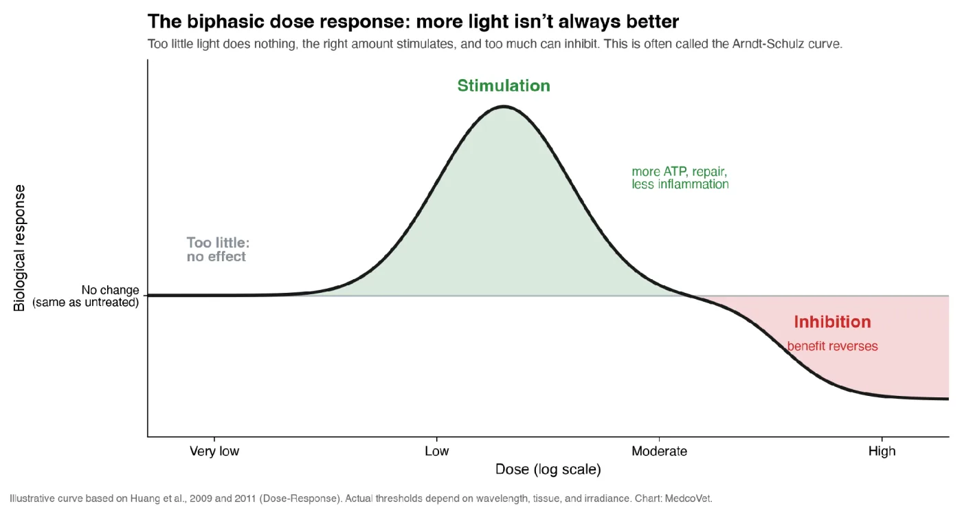
Figure 2. The biphasic dose response. Below a threshold, light has no effect. In the right range, it stimulates. Past that, the benefit reverses into inhibition. Illustrative curve based on Huang et al., 2009 and 2011. Chart: MedcoVet.
Researchers even use the inhibiting side on purpose. In lab studies, high-fluence red LED light slowed the growth, collagen production, and movement of skin fibroblasts, the cells that form scars (Mamalis et al., 2016). A follow-up trial tested 160 to 480 J/cm² on surgical scars. It didn’t meet its main goal, but scars improved more at some doses (Kurtti et al., 2021).
It isn’t only the total dose that behaves this way. Irradiance does too. The three-dimensional model below, from Michael Hamblin’s group at Harvard, shows that either irradiance or treatment time can push a treatment from stimulation into inhibition (Huang et al., 2011). It’s the turkey analogy in picture form: you need the right temperature and the right time, and too much of either ruins the result.
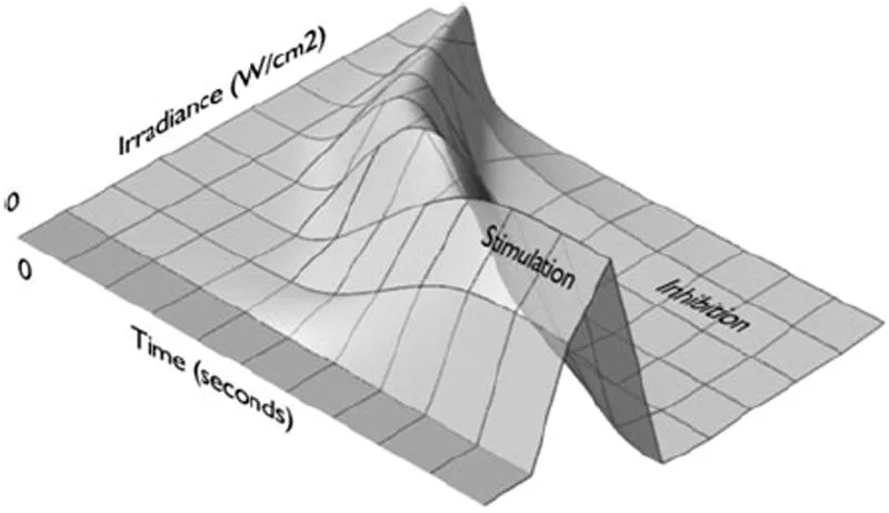
Figure 3. Three-dimensional model of the Arndt-Schulz curve, showing that either irradiance or illumination time can produce a biphasic response. Source: Huang YY, Sharma SK, Carroll J, Hamblin MR. Biphasic dose response in low level light therapy – an update. Dose-Response. 2011;9(4):602–618. © 2011 University of Massachusetts.
It doesn’t point to a single best solution; rather, cell culture, superficial wounds, arthritic joints, and nerve models differ in target biology, depth, wavelength, exposure schedule, and outcome measure. Moreover, even within a single condition, studies may use different devices and parameters.
Irradiance matters because it affects:
- How quickly photons arrive at the treatment surface
- How long the applicator must remain in place
- How sensitive the delivered exposure is to timing error
- The likelihood and rate of temperature rise
- How an uneven beam distributes exposure within the treatment area
- The starting amount of light available to travel into tissue
- Whether a published protocol can be reproduced
Complete biological exposure also depends on wavelength, pulse pattern, session frequency, total sessions, tissue condition, and how much light reaches and is absorbed by the target.
Two irradiance ranges, two different jobs
Here’s where red light therapy gets more interesting than a turkey. Your oven has two very different settings: bake, which cooks food all the way through, and broil, which blasts the surface with intense heat for a quick, different result. Irradiance has something similar: two broad ranges that do two different jobs.
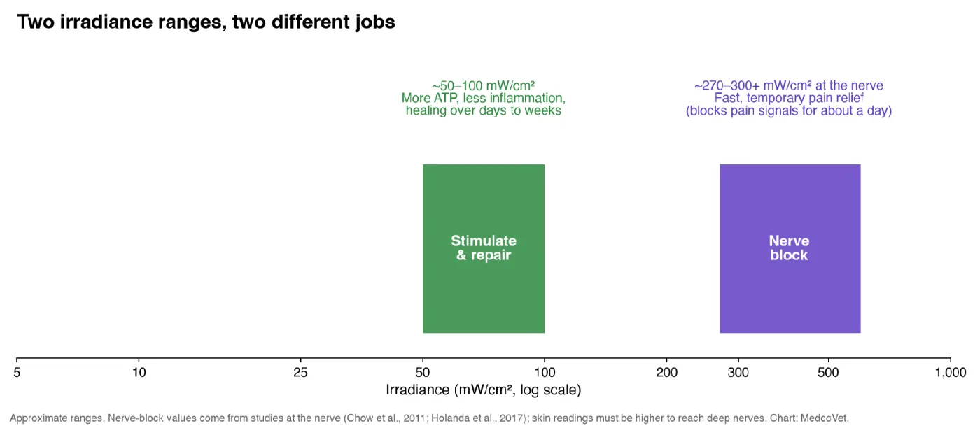
Figure 4. Lower irradiance stimulates cells and calms inflammation over time. Much higher irradiance at the nerve temporarily blocks pain signals. Approximate ranges. Chart: MedcoVet.
Lower irradiance: stimulate and repair
In this range, the light mainly acts on the mitochondria. It increases ATP, the cell’s energy supply, and sets off signals that calm inflammation and support repair (Chung et al., 2012). One study’s authors propose that lower irradiances, in the range of 10 to 100 mW/cm², may relieve pain by calming chronic inflammation (Holanda et al., 2017). This is the “bake” setting: the benefits build over repeated sessions, across days and weeks.
Much higher irradiance: temporary pain relief
At much higher irradiance, near-infrared light works differently. It temporarily slows the pain signals traveling along nerves. A systematic review of 44 studies found that laser light slowed nerve conduction and suppressed pain signals in many human and animal nerve models, likely by disrupting the internal “transport tracks” inside nerve fibers (Chow et al., 2011). At a high enough intensity, about 30% of nerve signaling is blocked within 10 to 20 minutes, and the effect wears off within about 24 hours (Cotler et al., 2015). In a rat model of nerve pain, 808 nm light at about 270 mW/cm², applied through the skin, reduced pain sensitivity within an hour, with effects fading within about 5 days (Holanda et al., 2017).
Other studies point the same way. In rats, 650 and 808 nm laser light applied over the sciatic nerve reduced nerve signals (Yan et al., 2011), and a review proposes that PBM pain relief may involve heat-stress and uncoupling-protein pathways (Cronshaw et al., 2019).
That’s fast, short-term pain relief, not healing. It’s the “broil” setting. The same researchers suggest combining the two: a high-irradiance treatment for quick relief, followed by lower-irradiance treatments to work on the inflammation behind the pain (Holanda et al., 2017).
Two cautions:
- The nerve-block numbers describe the light reaching the nerve. Light fades as it travels through skin, fur, and tissue, so 300 mW/cm² measured at the skin is far less at a nerve several millimeters down.
- High irradiance needs more care. It builds heat faster and shrinks the margin for timing errors (see “When irradiance is high” below). Like broiling, it’s not a set-it-and-walk-away setting.
So when you look at a device’s irradiance, ask which job it’s designed to do.
Is there an ideal irradiance for red light therapy?
For stimulation, the research range is wide, and many sources keep it below about 100 mW/cm²; some groups recommend under 100 mW/cm² at the target tissue (Zein et al., 2018). Sources commonly cite roughly 10 to 100 mW/cm². For contact devices on dogs and cats, which lose so much light to fur and tissue, 50 to 100 mW/cm² at the skin is a good working range for stimulating cells and calming inflammation. These numbers describe irradiance at the skin surface. The target tissue below gets much less, because skin, fat, and other tissue absorb and scatter the light on the way down.
WALT’s proposed veterinary guidelines, which The WAIT Test™ follows, give a range for each condition: 30 to 100 mW/cm² for wounds, 50 to 100 mW/cm² for most joints and the spine, and 100 to 250 mW/cm² for the hip, the deepest joint.
Below that range, the light can fall short of what cells need to respond. Researchers describe a threshold: if the irradiance is too low, there may be no response at all (Chung et al., 2012). It’s the turkey analogy again. Set the oven too low, and the bird never really cooks, no matter how long you leave it in. Above about 100 mW/cm², the opposite problems grow: more heat, less margin for timing errors, and a greater chance of tipping into the inhibiting side of the biphasic response.
Within that working range, there’s still no single best number. The right value depends on the details. One reason: tissues with many mitochondria, like muscle, brain, heart, and nerve, tend to respond to lower doses than tissues with fewer, like skin, tendon, and cartilage (Zein et al., 2018).
A useful value must be judged against:
- Target tissue and depth
- Wavelength or wavelength combination
- Desired biological effect
- Treatment area
- Continuous or pulsed operation
- Treatment time and frequency
- Patient anatomy, pigmentation, and coat
- Contact angle and pressure
- Heat management
- Evidence supporting the complete protocol
Simple rules such as ‘all PBM should be below X mW/cm²’ or ‘higher irradiance always penetrates more deeply’ reduce the entire treatment down to a single figure. Research shows that both dose and rate matter, but no single threshold fits every tissue or condition.
A better standard is:
Measure irradiance at the treatment surface. It needs to be high enough to deliver the right exposure in a reasonable time, but low enough to avoid overheating or timing problems. It should also match a protocol that fits the target tissue and the job you want the light to do.
When irradiance is very low
Very low irradiance is when the oven is set too low. At 5 mW/cm² at the skin surface, for example, the light may be enough for very superficial targets, but only a small fraction reaches deeper tissue, too little to count on. (In one widely cited study, 5 mW/cm² worked well, but that light was applied directly to the heart, not through skin (Oron et al., 2001).) And just like a turkey in a 100°F oven, leaving it on longer doesn’t fix the problem. Because PBM doesn’t reliably follow reciprocity, a long session at very low irradiance isn’t equivalent to a shorter one in the right range, even if the J/cm² adds up to the same number.
Very low irradiance also creates practical problems. Long sessions are hard to stick with: the user might stop early, move the applicator, lose skin contact, or treat areas inconsistently. Output can also drift over a long session as the battery drains or the device warms up.
Be especially cautious when a company pairs a very low irradiance with a long treatment time and a J/cm² figure. The math may add up, but the biology doesn’t.
When irradiance is high
High irradiance is the oven set too hot. Turning it up isn’t a shortcut to the same result in less time. A turkey roasted at 1,000°F for 72 minutes isn’t the same as one roasted at 400°F for 3 hours, even though the degree-hours match. Similarly, pushing irradiance above the stimulation range can shift cells from stimulation to inhibition (see Figure 3), so the benefit drops or reverses.
High irradiance also heats up faster and leaves less room for error. At 200 mW/cm², a 10-second error adds 2 J/cm² to the exposure. At 75 mW/cm², the same mistake changes it by 0.75 J/cm². Whether the extra heat matters depends on the wavelength, beam size, skin and coat, contact, cooling, movement, and exposure time.
One deliberate exception is much higher irradiance at the nerve (often hundreds of mW/cm²), used on purpose for short-term pain relief. That’s a different job, like switching the oven from bake to broil, and it needs a protocol and safety measures built for it.
What veterinary research tells us about irradiance
Veterinary PBM research does not identify one ideal irradiance for every animal and condition. It does show why reporting all parameters is essential.
A systematic review in 2023 examined 45 studies on laser therapy in animals, including 24 in dogs, one in cats, and the rest in horses. The authors observed conflicting results, substantial variation in wavelengths and treatment protocols, and a moderate or high risk of bias in several studies. They therefore stated that more definite guidelines are needed for clinical use and for higher-quality trials (Millis and Bergh, 2023).
Several individual studies show the range and the limits of comparison:
- A randomized study involving canine tibial plateau leveling osteotomies employed a dual-wavelength treatment ranging from 800 to 900 nm at a power level of 6 W over a stated area of 100 cm² and with a dose of 3.5 J/cm²; the simple average over the area amounts to about 60 mW/cm², although the use of a moving applicator and the nonuniformity of the field can cause local exposure to vary. The study found that the treated group had a higher peak vertical force at eight weeks (Rogatko et al., 2017).
- A study in dogs after thoracolumbar hemilaminectomy reported 810 nm treatment with 5.5 W/cm² and a very small stated beam area. It found no significant differences between groups in the main recovery measures. Do not compare the figure directly with a broad LED pad because beam size and application geometry differ (Bennaim et al., 2017).
- A retrospective investigation of dogs following surgery used synchronized emissions at 808 and 905 nm and recorded a dose of 4 J/cm² at six sites; although neurological scores improved in one subgroup, the time to start walking did not differ significantly. Because of its retrospective approach and varied treatment regimen, the study cannot draw causal conclusions (Bruno et al., 2020).
- The healthy-dog transmission study showed that a device’s incident output is only the first step. Coat, skin, wavelength, power, and shaving changed the amount detected after tissue transmission (Hochman-Elam et al., 2020).
- The equine skin study found that the tested devices did not rank by average power alone for transmitted energy (Luna et al., 2020).
These studies don’t mean you should always pick the highest irradiance or use the same number for every treatment. Instead, you should match the full published protocol to the animal, tissue, condition, and device.
Technical reference
Technical details
The sections above cover what most pet parents need to evaluate a device. The topics below go deeper into the physics and measurement details behind irradiance, for readers who want the full technical picture.
Thermal effects: PBM is not defined by a temperature reading alone
PBM is generally intended to produce photochemical or photophysical effects without relying on tissue heating as the main mechanism. That does not mean every PBM exposure is thermally neutral.
Absorbed optical energy can become heat. Surface temperature is affected by:
- Irradiance
- Wavelength and tissue absorption
- Beam area and spatial hot spots
- Treatment duration
- Continuous or pulsed output
- Skin pigmentation
- Fur and its color
- Contact pressure
- Stationary versus moving application
- Cooling and ambient conditions
An in vitro investigation into high-energy PBM settings in porcine muscle revealed wavelength-dependent temperature rises and indicated that moving the handpiece decreased heating. Although the study was carried out under specific laboratory conditions and therefore cannot establish a general safety limit, it shows that irradiance, beam size, wavelength, time, and movement must be considered together (Cronshaw et al., 2023). Further research on therapeutic lasers at 810 and 904 nm has also shown that wavelength and operating mode can produce different changes in skin temperature (Joensen et al., 2011).
A good consumer product should explain its thermal controls, maximum session time, proper use, and what to do if the area gets too warm. Just because the device feels cool doesn’t mean the tissue is safe, and feeling warmth doesn’t guarantee you’ve reached the right PBM dose.
Does higher irradiance send light deeper?
Not necessarily.
For a given wavelength, beam shape, and path through the tissue, increasing surface irradiance will increase the amount of light available at depth. If 1% of the incident light reaches the target, then increasing the incident light will increase the transmitted light proportionally, provided the optical conditions remain the same.
But irradiance does not determine the fraction that penetrates. That fraction is strongly affected by:
- Wavelength
- Tissue composition and thickness
- Melanin, blood, and water absorption
- Scattering
- Beam diameter and divergence
- Contact and compression
- Fur or other barriers
- Applicator angle
Higher surface irradiance means more light hits the surface, but it doesn’t automatically mean better penetration or a guaranteed dose deeper in the tissue.
An equine skin study illustrates the limit of the shortcut. In that head-to-head comparison, the tested superpulsed device transmitted a higher percentage of its output through skin than the tested Class IV device (absolute transmitted power wasn’t compared). Because the devices differed in several ways, the result does not prove that lower power is generally superior. It does show that device output alone cannot predict transmission (Luna et al., 2020).
Surface irradiance is not target-tissue irradiance
Stating that a joint several centimeters below the skin surface receives 75 mW/cm² when the product states 75 mW/cm² at the skin would be incorrect. Reflection, scattering, absorption, and geometry alter the field inside the body.
Determining irradiance inside tissue may require:
- Direct detector measurements in or behind tissue
- Tissue phantoms with known optical properties
- Monte Carlo or other optical modeling
- Anatomical imaging and tissue-thickness estimates
- Measurements specific to the wavelength, applicator, and patient type
Even the most advanced models have to make assumptions. When giving product specifications, clearly state where the measurement was taken and avoid turning a surface reading into an unsubstantiated claim about the internal dose.
The same caution applies to studies. An irradiance reported “at the target” may mean the skin surface over the anatomical target, not the internal structure itself. Read the methods.
Why distance changes irradiance
The farther a light source is from a surface, the larger the area its output spreads over, and the lower the irradiance. When the treatment plane is close, you will see individual bright spots with gaps, but when the plane is farther away, the light is more uniform, although at a lower average irradiance.
A specification should therefore state:
- Exact measurement distance
- Distance from which the reference point was taken, such as lens, housing, or spacer
- Treatment area at that distance
- Irradiance map at that distance
- Recommended treatment distance
- Whether a cover, spacer, or lens was in place
Why the inverse-square law is often misapplied
The inverse-square law describes irradiance from an ideal point source in free space. A large nearby panel, a lens-controlled LED, a cluster of overlapping emitters, and a contact applicator are not ideal point sources at typical treatment distances.
Increasing the distance from a real panel does not ensure that the irradiance will be exactly one-quarter. For the device in question, you must measure the change. Recent studies of multi-LED systems have used two-dimensional maps at known distances because emitter overlap and detector angle can materially affect the result (Gerelli et al., 2026).
Don’t use a contact measurement to figure out a treatment done several inches away. Also, don’t rely on the inverse-square law if the manufacturer can give you a direct measurement.
Angle, curvature, and movement also matter
An irradiance value measured on a flat surface with the applicator perpendicular to it may not describe treatment on a curved shoulder, hip, paw, or spine.
When light strikes at an angle:
- The same beam can cover a larger projected area
- Irradiance per unit of surface can fall
- Reflection can increase
- One edge may be closer to the source than another
Curved surfaces can affect contact and create gaps. A stiff panel might be close in one area but farther away in another. A flexible pad fits better but can still vary depending on emitter spacing and applied pressure.
Movement changes the picture again. Although scanning a beam can distribute total energy over a longer path, it is difficult to determine dwell time at any given point unless you carefully control the speed, overlap, path, and number of repetitions. A stationary spot specification cannot be directly applied to a scanning procedure.
Peak, average, and spatial irradiance are different
The word peak can refer to two different things:
- Temporal peak irradiance: the highest irradiance while a pulse is on.
- Spatial peak irradiance: the highest point within a beam or LED array.
These are not the same quantity.
An LED panel might run all the time but have a peak right under one light. A pulsed laser could have a high peak during each pulse, but the time-averaged value is much lower. If a spec just says ‘peak irradiance,’ it’s unclear unless it explains what kind of peak it means.
For treatment calculations, ask for:
- Time-averaged, area-averaged irradiance
- Temporal peak irradiance for pulsed modes
- Spatial peak and minimum within the stated treatment area
- The method used to calculate each value
For a pulsed device, use the time-averaged irradiance for dose math. The Average Power guide explains peak versus average output and duty cycle in full.
Spatial uniformity: one number can hide the treatment area
Real treatment fields are rarely perfectly uniform.
- A laser beam may have a Gaussian profile, with a bright center and weaker edges.
- An LED array may have peaks under each emitter and lower values between them.
- Optical lenses may create rings or edge falloff.
- Mixed-wavelength arrays may distribute each wavelength differently.
- A close treatment distance may increase local variation.
Suppose a 20 cm² pad reports an average of 50 mW/cm². That average could describe a fairly even field from 45 to 55 mW/cm². It could also hide small 200 mW/cm² hot spots and large weak areas. The average alone cannot distinguish them.
There’s some nuance. Light scatters sideways under the skin, so small gaps between closely spaced emitters partly fill in below the surface. Large gaps and hot spots still matter. Beam shape matters too: a flat-top beam produced more even effects on isolated mitochondria across the treated area than standard Gaussian probes, whose light is concentrated in the center (Amaroli et al., 2021).
A useful irradiance map should report:
- The mapped area and grid spacing
- Measurement distance and orientation
- Average, median, maximum, and minimum
- Variation or uniformity statistic
- Each wavelength separately when their spatial patterns differ
- The device mode and stabilization time
- Whether interpolation or smoothing was used
Hadis and his colleagues demonstrated that laser and LED sources can have very different spatial profiles and emphasized the importance of measuring the area of the beam at the treatment surface rather than assuming it is the same as the aperture (Hadis et al., 2016). In a later study involving an LED array, sources covering the wavelength range from 400 to 850 nm were examined, and in this in vitro study of human dental pulp cells, the biological response depended on the wavelength, the exposure time, and when the assessment was made (Hadis et al., 2017). A multi-panel study in 2026 reached a similar conclusion, showing that biological responses varied with spatial irradiance across the entire device field (Gerelli et al., 2026).
The point isn’t that every variation makes a device useless. The reported number should reflect what the treatment area actually receives.
Beam area must be defined at the treatment plane
Irradiance calculations are only as accurate as the area measurement.
The relevant area is not necessarily:
- The physical size of the device head
- The lens diameter
- The aperture opening
- The area covered by LED housings
- The manufacturer’s named “coverage area”
It is the illuminated beam or treatment area at the specified treatment plane, defined by a stated method.
Beam edges are not always sharp. For Gaussian and irregular beams, different conventions can produce different area values. A study or company should state how the boundary was chosen and whether the calculation used an effective beam area, a threshold contour, a diameter at a specified intensity, or another method.
This is not a minor detail. If a 1 W beam is said to cover 1 cm², the calculated irradiance is 1 W/cm². If measurement shows the effective area is actually 2 cm², the area-average value is 0.5 W/cm². Area error passes directly into irradiance error.
A study of 24 clinical PBM laser devices found large differences between the stated and actual power and beam diameter. Nine devices had measured power within 90% to 110% of the nominal value, and measured beam diameters varied from 38% to 543% of the stated figures (Girasol et al., 2022).
Multi-wavelength devices need more than one combined figure
A device that emits 660 and 850 nm might report a combined irradiance of 75 mW/cm². That number does not reveal whether the split is:
- 37.5 mW/cm² at each wavelength
- 60 mW/cm² at 660 nm and 15 mW/cm² at 850 nm
- Another ratio that changes by mode or position
Because wavelength affects absorption, scattering, photon energy, and biological relevance, report each channel separately and in combination.
Ask for:
- Irradiance by wavelength at the treatment plane
- Combined area-average irradiance
- Spatial map for each wavelength
- Which wavelengths are active in each mode
- Whether the detector was calibrated for every emitted band
- Whether simultaneous operation changes either channel’s output
It’s fine to add channel irradiances if they’re measured over the same area, at the same distance, and in the same mode. Don’t add values taken under different conditions.
Irradiance is an energy rate, not a photon count
A watt measures energy used per second, and it doesn’t track photons directly.
The energy of one photon is:
Photon energy = hc / wavelength
Photons with a longer wavelength have less energy each. And so, at the same irradiance, 850 nm light has more photons per second per square centimeter than 660 nm light. However, this doesn’t mean 850 nm is automatically more effective; tissue transmission, absorption by relevant molecules, and the biological response all vary with wavelength.
For most product comparisons, it’s easier to use measured irradiance by wavelength instead of photon count. Still, remember that the same mW/cm² at different wavelengths doesn’t mean the same number of photons or the same effect in tissue.
Laser and LED irradiance cannot be compared by one number alone
Irradiance has the same physical unit for lasers and LEDs, but the exposure field can differ in several ways:
- Beam profile and divergence
- Spectral bandwidth
- Coherence
- Pulse structure
- Spot size
- Contact method
- Spatial uniformity
- Movement during treatment
A narrow laser spot at 100 mW/cm² and a broad LED pad at 100 mW/cm² have the same area-rate unit, but they may treat very different areas and use different protocols. A point applicator may be moved or placed at several sites. A pad may expose the full region at once.
Just because a laser protocol works doesn’t mean an LED device with the same irradiance will too. For the evidence to apply, the wavelength, irradiance pattern, time, pulse mode, area, use, target, and session schedule must be similar.
How irradiance should be measured
Estimate irradiance using optical equipment calibrated for the wavelength, beam shape, power range, and pulse characteristics.
Common approaches include:
- Measuring total optical power and dividing it by a properly measured beam area
- Using a calibrated irradiance detector with a known active area
- Scanning a small detector across a grid to build a two-dimensional map
- Using a camera-based beam profiler calibrated for spatial power distribution
- Using a spectroradiometer when wavelength-resolved irradiance is required
- Using an integrating sphere for total output, paired with a separate beam-area or spatial measurement
No one instrument works perfectly for every light source. A detector can cause errors if:
- Its spectral response does not match the emitted wavelengths
- Its active area is too large to resolve hot spots
- Its active area is too small to collect the whole beam when total power is intended
- It saturates at peak power
- It is too slow to resolve pulses
- It lacks cosine correction for light arriving over a wide range of angles
- Its calibration is expired or does not cover the measured range
- It is positioned at the wrong distance or angle
What a useful measurement statement should include
A credible specification should disclose:
- Device model and production version
- Active wavelength or wavelengths
- Operating mode and power setting
- Continuous or pulsed output
- Warm-up or stabilization procedure
- Measurement plane and distance
- Treatment or beam area at that plane
- Instrument manufacturer and model
- Detector type, active area, and spectral range
- Calibration date or traceability
- Sampling grid or beam-area method
- Average, peak, minimum, and variation across the area
- Number of devices and repeat measurements
- Manufacturing tolerance
- Output change during the recommended session
Independent testing can help build trust, but ‘third-party tested’ doesn’t mean much unless you know the test conditions and results.
Why lux, lumens, and phone apps cannot replace irradiance
Lux and lumens account for the human eye’s sensitivity to visible light; however, PBM devices usually emit near-infrared wavelengths that aren’t visible to the human eye, and lux meters aren’t built to measure them accurately.
A red device that looks bright might not have much optical power. A near-infrared device can look dim or even dark but still give off a lot of energy. So, you can’t judge by how bright it looks.
Phone cameras and light-meter apps also aren’t suitable for validating PBM irradiance. Their spectral filters, automatic exposure controls, unknown calibration, sensor saturation, and limited near-infrared response prevent a trustworthy mW/cm² measurement.
That includes inexpensive “solar power meters,” the most common consumer meter. They’re calibrated for sunlight, and a detector’s response changes with wavelength (Hadis et al., 2016). In one light-panel company’s comparison against a reference measurement, a solar meter read three panels 31% to 88% too high (GembaRed).
Use radiometric equipment that’s calibrated for your light source. Don’t rely on photometric units or just how the light looks.
Measured output matters more than component ratings
An irradiance estimate built by multiplying LED count by each diode’s catalog rating can fail for several reasons:
- Electrical drive current may differ from the rating condition
- Optical efficiency varies with temperature
- Lenses change the illuminated area and overlap
- Driver losses affect output
- Enclosure windows absorb some light
- Diode output varies between production lots
- Multiple wavelengths may have different efficiencies
- Component ratings may be maxima rather than normal operating values
Test the finished device in its actual treatment mode and at the treatment surface. Also, check it after it’s warmed up, since the output at startup might be different from what you get after a few minutes.
The Girasol study serves as a practical warning: the stated values for power and beam were often different from those obtained by direct measurement, which means the calculated irradiance and dose are also likely to be wrong (Girasol et al., 2022).
FAQ
Frequently asked questions about irradiance
What is a good irradiance for red light therapy?
There is not a single figure that applies to all cases. Research points to two broad ranges with different jobs: roughly 50 to 100 mW/cm² to stimulate cells and calm inflammation, and much higher irradiance, often hundreds of mW/cm² at the nerve, for temporary pain relief. Within those, a useful irradiance must provide an acceptable surface exposure in a reasonable time, keep heating under control, and suit the wavelength, target tissue, area, pulse mode, and treatment schedule. Compare the entire protocol rather than just one number.
Is 100 mW/cm² too high?
Not for stimulation. It’s the top of the 50 to 100 mW/cm² working range. Above it, heat builds faster, and the risk of tipping into inhibition grows. At 100 mW/cm², the surface exposure builds up at 0.1 J/cm² every second, so accurate timing and temperature control matter. For a deep joint like the hip, WALT’s guidelines go up to 250 mW/cm².
Is 10 mW/cm² too low?
For contact devices on pets, usually yes. Measured at the skin, it’s at the low end of the commonly cited 10 to 100 mW/cm² range and below the 50 to 100 mW/cm² WALT suggests for most joints. It also falls short of the roughly 11 mW/cm² of usable light The WAIT Test™ asks for at the skin over most joints, before any coat losses. Like a turkey in an oven set too low, a longer session doesn’t make up the difference, because PBM doesn’t reliably follow reciprocity.
Can high irradiance relieve pain?
Yes, in a specific way. At much higher irradiance, often hundreds of mW/cm² at the nerve, near-infrared light can temporarily slow pain signals, with the effect wearing off within about a day. That’s short-term relief, not healing, and the light must actually reach the nerve at that intensity.
Is irradiance the same as power density?
Power density is the common PBM term for irradiance. Irradiance is the more precise radiometric term for radiant power arriving per unit area.
Is irradiance the same as energy density?
No. Irradiance is measured in W/cm². Energy density, more precisely radiant exposure, is measured in J/cm². Multiply irradiance by time to calculate radiant exposure.
Can I calculate irradiance from watts?
Only if you know the optical power arriving at the treatment plane and the illuminated area at that same plane. The simple calculation also assumes a useful definition of area and produces an average, not a spatial map.
Can I figure out treatment time using irradiance?
Yes, if you have a justified target radiant exposure: Time = radiant exposure/irradiance. The equation does not tell you what radiant exposure is appropriate. That must come from a protocol relevant to the tissue, condition, wavelength, and treatment schedule.
Will a higher irradiance result in a shorter treatment?
Mathematically, yes, for the same J/cm² over the same area. Biologically, they aren’t equivalent. PBM doesn’t reliably follow reciprocity, and heat, timing, and pulse structure all change with the rate. Think of the turkey: hotter and shorter isn’t the same dinner.
Does higher irradiance go further?
When all the other conditions are unchanged, it can increase the amount of light available at greater depths. However, it does not increase the fraction transmitted, nor does it guarantee that a deep target receives an effective dose; wavelength, tissue, beam geometry, contact, and barriers all play a role.
What causes irradiance to decrease as the distance increases?
The light typically covers a wider area, and in the case of an LED array the beams also overlap differently. The precise relationship varies with the shape of the light source, which is why you should use measurements taken at the recommended distance rather than assuming an inverse-square law applies for a point source.
Is the center reading the irradiance?
It is the irradiance at the center measurement point. It may be a spatial peak and should not be presented as the average for the whole treatment area unless mapping supports that conclusion.
How is average irradiance different from peak irradiance?
Average irradiance can refer to time averaging, area averaging, or both. Temporal peak is the maximum during a pulse. Spatial peak is the maximum point in the field. A company should define each term.
Can a lux meter be used to measure the irradiance of red light therapy?
Lux accounts for how the human eye responds and does not accurately measure near-infrared radiation. For this purpose, you should use radiometric equipment that has been calibrated for the wavelengths being emitted.
Can two devices that have the same irradiance provide different treatments?
Certainly. They can vary in wavelength, uniformity, pulse structure, spot size, contact, treatment time, spectral bandwidth, output stability, and the total area being treated.
Does pulsing make high peak irradiance more effective?
There is no universal answer. Pulse frequency, width, duty cycle, peak and average output, wavelength, and target all matter. Peak irradiance alone cannot establish dose or effectiveness.
What amount of irradiance reaches a joint?
A surface specification can’t answer that. To determine internal irradiance, you need transmission measurements or verified modeled data for the wavelength, anatomy, tissue depth, coat, and application geometry.
What is the relevance of fur when the open-air irradiance is high?
Fur can block or divert light away from the skin. Because this effect depends on the coat type and its location, a high open-air figure does not guarantee skin exposure. What matters is reliable skin access.
Should irradiance be measured through a spacer or cover?
If you use a spacer or cover during treatment, measure the most relevant value with it in place at the surface where the skin will be. Materials, distance, and angle can all change delivery.
The bottom line
Irradiance is the oven temperature of red light therapy. It tells you how concentrated the light is on each square centimeter of a specified treatment surface, and it links the measured optical power and the beam area to the treatment time and the radiant exposure.
Irradiance is essential, but it’s not a single score that tells you how effective a device is. Remember the two ranges: lower irradiance to stimulate and repair over time, and much higher irradiance at the nerve for short-term pain relief.
A credible device should report time-averaged, area-averaged irradiance at the actual treatment plane. It should state the distance, area, mode, wavelengths, pulse settings, measurement method, spatial variation, output stability, and tolerance. Its irradiance and treatment time should reproduce the claimed J/cm².
Like an oven, irradiance needs to be in the right range. If it’s too low, more time won’t make up for it. Too high, and you don’t get the same result faster: you risk inhibition, extra heat, and bigger timing errors. Because PBM doesn’t reliably follow reciprocity, equal J/cm² delivered at different irradiances does not produce the same biological response.
The best irradiance isn’t just the biggest number. It’s the measured and controlled rate that fits a complete, practical protocol for the tissue you want to treat.
Continue The WAIT Test™
This guide covers Irradiance, the third of the four components. To run the full evaluation, including the data you need and the decision rules, use The WAIT Test™ guide. The other three components:
- Wavelengths: Is it the right kind of light for the target tissue?
- Average Power: Is there enough light to treat the whole area?
- Target Tissue: Does the light reach the target tissue?
The four components work together. Passing one does not compensate for missing information in another.
The WAIT Test™ was developed by MedcoVet. The framework may be shared for educational purposes when its four components remain unchanged, and MedcoVet is credited as its creator.
The WAIT Test™ is an educational framework for comparing device specifications. It does not determine whether PBM is appropriate for a particular pet, diagnosis, or treatment site. Make treatment decisions with a qualified veterinary professional.
For every WAIT guide and tool in one place, start at The WAIT Test™ hub.
Ready to take the next step?
Book a free consultation with a licensed clinician to discuss your pet’s condition and treatment plan.
Spock
Learn more about the MedcoVet Luma and how it is designed for at-home red light therapy for pets.
Sources
Research and references
- Jenkins PA, Carroll JD. How to report low-level laser therapy (LLLT)/photomedicine dose and beam parameters in clinical and laboratory studies. Photomedicine and Laser Surgery. 2011;29(12):785-787. PubMed
- Hadis MA, Zainal SA, Holder MJ, et al. The dark art of light measurement: accurate radiometry for low-level light therapy. Lasers in Medical Science. 2016;31(4):789-809. PubMed
- Girasol CE, Braz GdA, Bachmann L, Celli J, Guirro RRdJ. Laser light sources for photobiomodulation: the role of power and beam characterization in treatment accuracy and reliability. PLOS ONE. 2022;17(3). Full text
- Chung H, Dai T, Sharma SK, Huang YY, Carroll JD, Hamblin MR. The nuts and bolts of low-level laser (light) therapy. Annals of Biomedical Engineering. 2012;40(2):516-533. PubMed
- Lanzafame RJ, Stadler I, Kurtz AF, et al. Reciprocity of exposure time and irradiance on energy density during photoradiation on wound healing in a murine pressure ulcer model. Lasers in Surgery and Medicine. 2007;39(6):534-542. PubMed
- Almeida-Lopes L, Rigau J, Zangaro RA, Guidugli-Neto J, Jaeger MM. Comparison of low-level laser therapy effects on cultured human gingival fibroblasts proliferation using different irradiance and same fluence. Lasers in Surgery and Medicine. 2001;29(2):179-184. PubMed
- Huang YY, Chen AC, Carroll JD, Hamblin MR. Biphasic dose response in low level light therapy. Dose-Response. 2009;7(4):358-383. Full text
- Huang YY, Sharma SK, Carroll J, Hamblin MR. Biphasic dose response in low level light therapy: an update. Dose-Response. 2011;9(4):602-618. Full text
- Sharma SK, Kharkwal GB, Sajo M, et al. Dose-response effects of 810 nm laser light on mouse primary cortical neurons. Lasers in Surgery and Medicine. 2011;43(8):851-859. PubMed
- Chow R, Armati P, Laakso EL, Bjordal JM, Baxter GD. Inhibitory effects of laser irradiation on peripheral mammalian nerves and relevance to analgesic effects: a systematic review. Photomedicine and Laser Surgery. 2011;29(6):365-381. PubMed
- Cotler HB, Chow RT, Hamblin MR, Carroll J. The use of low level laser therapy (LLLT) for musculoskeletal pain. MOJ Orthopedics and Rheumatology. 2015;2(5):00068. Full text
- Holanda VM, Chavantes MC, Wu X, Anders JJ. The mechanistic basis for photobiomodulation therapy of neuropathic pain by near infrared laser light. Lasers in Surgery and Medicine. 2017;49(5):516-524. PubMed
- Zein R, Selting W, Hamblin MR. Review of light parameters and photobiomodulation efficacy: dive into complexity. Journal of Biomedical Optics. 2018;23(12):1-17. PubMed
- Hashmi JT, Huang YY, Sharma SK, et al. Effect of pulsing in low-level light therapy. Lasers in Surgery and Medicine. 2010;42(6):450-466. PubMed
- Hadis MA, Cooper PR, Milward MR, et al. Development and application of LED arrays for use in phototherapy research. Journal of Biophotonics. 2017;10(11):1514-1525. PubMed
- Gerelli E, Wagnières G, Joniová J. Radiometric and spectral characterization of multipanel planar light-emitting devices: a generalizable irradiance mapping approach for dose estimation. Journal of Biomedical Optics. 2026;31(5):055001. PubMed
- Cronshaw M, Parker S, Grootveld M, Lynch E. Photothermal effects of high-energy photobiomodulation therapies: an in vitro investigation. Biomedicines. 2023;11(6):1634. PubMed
- Joensen J, Demmink JH, Johnson MI, Iversen VV, Lopes-Martins RAB, Bjordal JM. The thermal effects of therapeutic lasers with 810 and 904 nm wavelengths on human skin. Photomedicine and Laser Surgery. 2011;29(3):145-153. DOI
- Hochman-Elam LN, Heidel RE, Shmalberg JW. Effects of laser power, wavelength, coat length, and coat color on tissue penetration using photobiomodulation in healthy dogs. Canadian Journal of Veterinary Research. 2020;84(2):131-137. PubMed
- Luna SPL, Schoen A, Trindade PHE, da Rocha PB. Penetration profiles of a Class IV therapeutic laser and a photobiomodulation therapy device in equine skin. Journal of Equine Veterinary Science. 2020;85:102846. PubMed
- Millis DL, Bergh A. A systematic literature review of complementary and alternative veterinary medicine: laser therapy. Animals. 2023;13(4):667. PubMed
- Rogatko CP, Baltzer WI, Tennant R. Preoperative low level laser therapy in dogs undergoing tibial plateau levelling osteotomy: a blinded, prospective, randomized clinical trial. Veterinary and Comparative Orthopaedics and Traumatology. 2017;30(1):46-53. PubMed
- Bennaim M, Porato M, Jarleton A, et al. Preliminary evaluation of the effects of photobiomodulation therapy and physical rehabilitation on early postoperative recovery of dogs undergoing hemilaminectomy for treatment of thoracolumbar intervertebral disk disease. American Journal of Veterinary Research. 2017;78(2):195-206. PubMed
- Bruno E, Canal S, Antonucci M, et al. Perilesional photobiomodulation therapy and physical rehabilitation in post-operative recovery of dogs surgically treated for thoracolumbar disk extrusion. BMC Veterinary Research. 2020;16:120. PubMed
- Avci P, Gupta A, Sadasivam M, et al. Low-level laser (light) therapy (LLLT) in skin: stimulating, healing, restoring. Seminars in Cutaneous Medicine and Surgery. 2013;32(1):41-52. Full text
- Barolet D, Christiaens F, Hamblin MR. Infrared and skin: friend or foe. Journal of Photochemistry and Photobiology B. 2016;155:78-85. Full text
- Zeng Y, et al. Can transcranial photobiomodulation improve cognitive function in TBI patients? A systematic review. Frontiers in Psychology. 2024;15:1378570. Full text
- Yan W, Chow R, Armati PJ. Inhibitory effects of visible 650-nm and infrared 808-nm laser irradiation on somatosensory and compound muscle action potentials in rat sciatic nerve: implications for laser-induced analgesia. Journal of the Peripheral Nervous System. 2011;16(2):130-135. PubMed
- Cronshaw M, Parker S, Arany P. Feeling the heat: evolutionary and microbial basis for the analgesic mechanisms of photobiomodulation therapy. Photobiomodulation, Photomedicine, and Laser Surgery. 2019;37(9):517-526. PubMed
- Mamalis A, Koo E, Garcha M, Murphy WJ, et al. High fluence light emitting diode-generated red light modulates characteristics associated with skin fibrosis. Journal of Biophotonics. 2016;9(11-12):1167-1179. PubMed
- Kurtti A, Nguyen JK, Weedon J, Mamalis A, et al. Light emitting diode-red light for reduction of post-surgical scarring: results from a dose-ranging, split-face, randomized controlled trial. Journal of Biophotonics. 2021;14(7):e202100073. PubMed
- Amaroli A, Arany P, Pasquale C, Benedicenti S, et al. Improving consistency of photobiomodulation therapy: a novel flat-top beam hand-piece versus standard Gaussian probes on mitochondrial activity. International Journal of Molecular Sciences. 2021;22(15):7788. PubMed
- Haslerud S, et al. Achilles tendon penetration for continuous 810 nm and superpulsed 904 nm lasers before and after ice application: an in situ study on healthy young adults. Photomedicine and Laser Surgery. 2017;35(10):567-575. PubMed
- Oron U, Yaakobi T, Oron A, et al. Low-energy laser irradiation reduces formation of scar tissue after myocardial infarction in rats and dogs. Circulation. 2001;103(2):296-301. PubMed
- GembaRed. What is the real intensity of my LED panel? The dark deception of red light therapy. Full text


