Elbow Dysplasia in Dogs
What it is, what it looks like, signs to watch for, and treatment options that support comfort and mobility
Help your dog move with less elbow pain
Talk with a MedcoVet clinician about your dog’s limp, stiffness, elbow discomfort, and at-home support options.
Medically reviewed by: Kristy Williams, CVT, CCFT (Specialties: Pet rehabilitation, pain management, photobiomodulation)
Reviewed: [June 2026]
Updated: [June 2026]
What is elbow dysplasia in dogs?
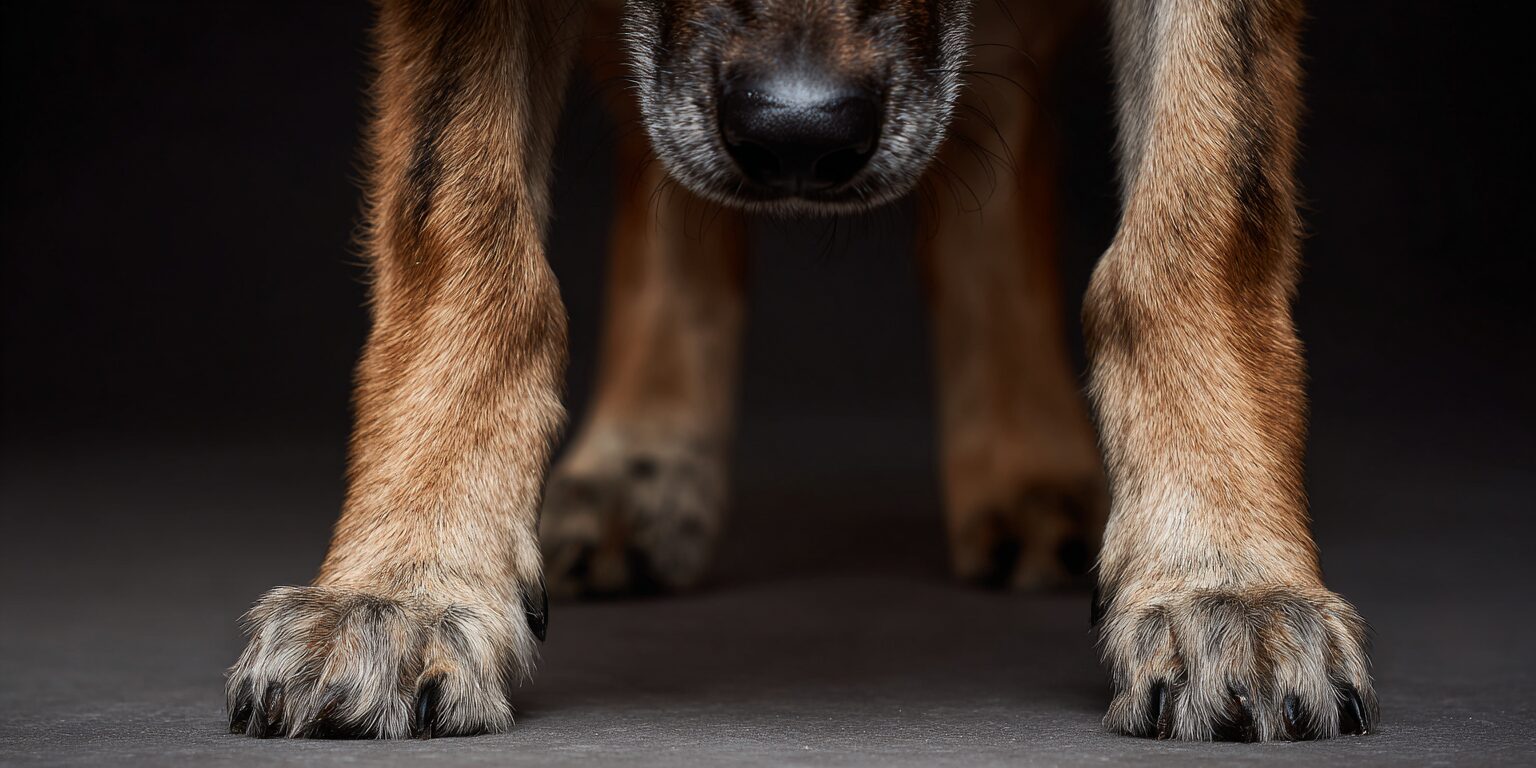
Elbow dysplasia in dogs is a developmental joint condition where the dog’s elbow joint does not form or move correctly. The canine elbow is a complex joint made of three bones: the humerus, radius, and ulna. Abnormal development of the bones and cartilage in the dog’s elbow joint can lead to joint misalignment and instability. When these bones do not fit together smoothly, the joint surface can become damaged, causing joint pain, forelimb lameness, cartilage wear, joint swelling, joint instability, and degenerative joint disease.
If your dog is limping, stiff, or showing signs of elbow pain:
Want help thinking through your dog’s symptoms?
Sprite
Elbow dysplasia is the second-most common cause of elbow lameness in dogs, with osteoarthritis being the first.
Why elbow dysplasia happens
Canine elbow dysplasia is one of the most common causes of front-leg lameness, especially in young, rapidly growing large-breed dogs. Symptoms often appear between 5 and 18 months of age, though some dogs are diagnosed later after arthritis or chronic pain becomes more obvious.
Elbow dysplasia is considered a multifactorial condition. That means several problems can contribute to the same painful result.
It may involve one or more of the following:
- Fragmented coronoid process
- Fragmented medial coronoid process
- Medial coronoid process disease
- Ununited anconeal process, or UAP
- Osteochondrosis or osteochondritis dissecans (OCD) of the medial humeral condyle
- Cartilage flap formation
- Elbow incongruity
- Joint incongruity
- Abnormal bone growth
- Medial compartment disease
That sounds like a lot. Here is the simple version.
The elbow joint needs three bones to fit together precisely. If one part grows unevenly, if cartilage does not develop normally, or if one joint surface takes too much pressure, the affected joint can become painful and unstable.
Over time, joint trauma inside the elbow can lead to cartilage erosion, cartilage loss, joint degeneration, and arthritis.
Which dogs are most at risk?

Elbow dysplasia has a strong suspected genetic component. It is especially common in certain large breeds.
Commonly affected breeds include:
- Labrador Retrievers
- Golden Retrievers
- German Shepherd Dogs
- Rottweilers
- Bernese Mountain Dogs
- Newfoundland dogs
- Mastiffs
- Other large and giant breed dogs
The hereditary basis of conditions like ununited anconeal process and medial coronoid disease has been suggested, though the inheritance pattern is not always simple.
Growth matters too.
Puppies that grow too quickly, often from calorie-dense puppy food, can place too much mechanical stress on developing joints. Improper dietary balance of calcium and phosphorus can also disrupt normal cartilage development.
Screening breeding dogs through health testing can help reduce elbow dysplasia in future litters. Parents with “normal” elbow grading are less likely to produce dysplastic puppies than affected parents.
You cannot prevent every case. You can reduce risk with responsible breeding, proper puppy nutrition, healthy growth, and veterinary guidance.
Signs of elbow dysplasia in dogs
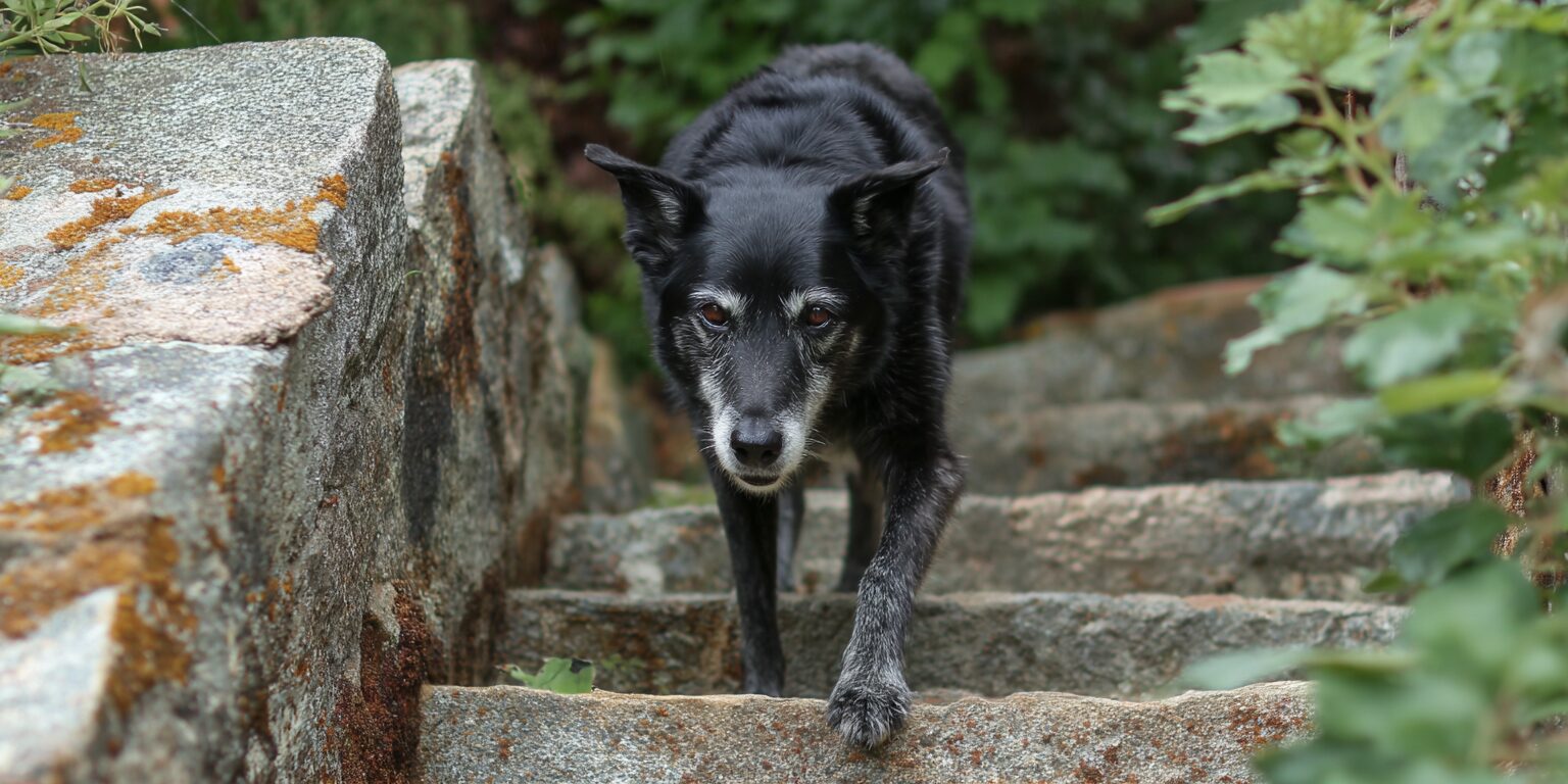
Signs of elbow dysplasia in dogs often start as a front-leg limp. Sometimes it is subtle. Sometimes it shows up only after exercise. Affected dogs may exhibit a range of symptoms and behavioral changes, such as less interest in physical activities due to pain and stiffness.
Common clinical signs include:
- Front leg lameness
- Stiffness upon rising
- Abnormal leg positioning
- Head bobbing while walking
- Reduced range of motion
- Joint swelling
- Exercise reluctance
- Limping in one or both front legs
- Swollen elbows
- Holding the elbow at an unusual angle
- Turning the paw outward
- Pain when the elbow is touched
- Muscle atrophy in the affected leg / shoulder
- Shortened stride
- Difficulty getting up
- Less interest in play or walks
Dogs with elbow dysplasia may limp more after exercise because movement increases joint discomfort. They may also hold their elbows out or shift weight to reduce pressure on the painful joint.
In severe cases, the elbow may look swollen or puffy due to inflammation or fluid buildup.
If your dog has a front-leg limp that keeps coming back, do not brush it off. Early diagnosis can protect long-term joint health.
What does elbow dysplasia look like in dogs?
Elbow dysplasia can look like a dog who is “just a little stiff,” especially at first.
You may notice your dog:
- Takes shorter steps with one front leg
- Bobs their head when walking
- Avoids running
- Gets worse after play
- Looks stiff when rising
- Holds one front leg slightly rotated
- Licks near the elbow
- Hesitates before jumping down
- Walks unevenly on hard surfaces
- Has less enthusiasm for walks
Head bobbing is a common clue. Dogs often lift the head when the painful front leg touches the ground and lower it when the comfortable leg lands.
It is subtle once you know what to look for.
How veterinarians diagnose elbow dysplasia
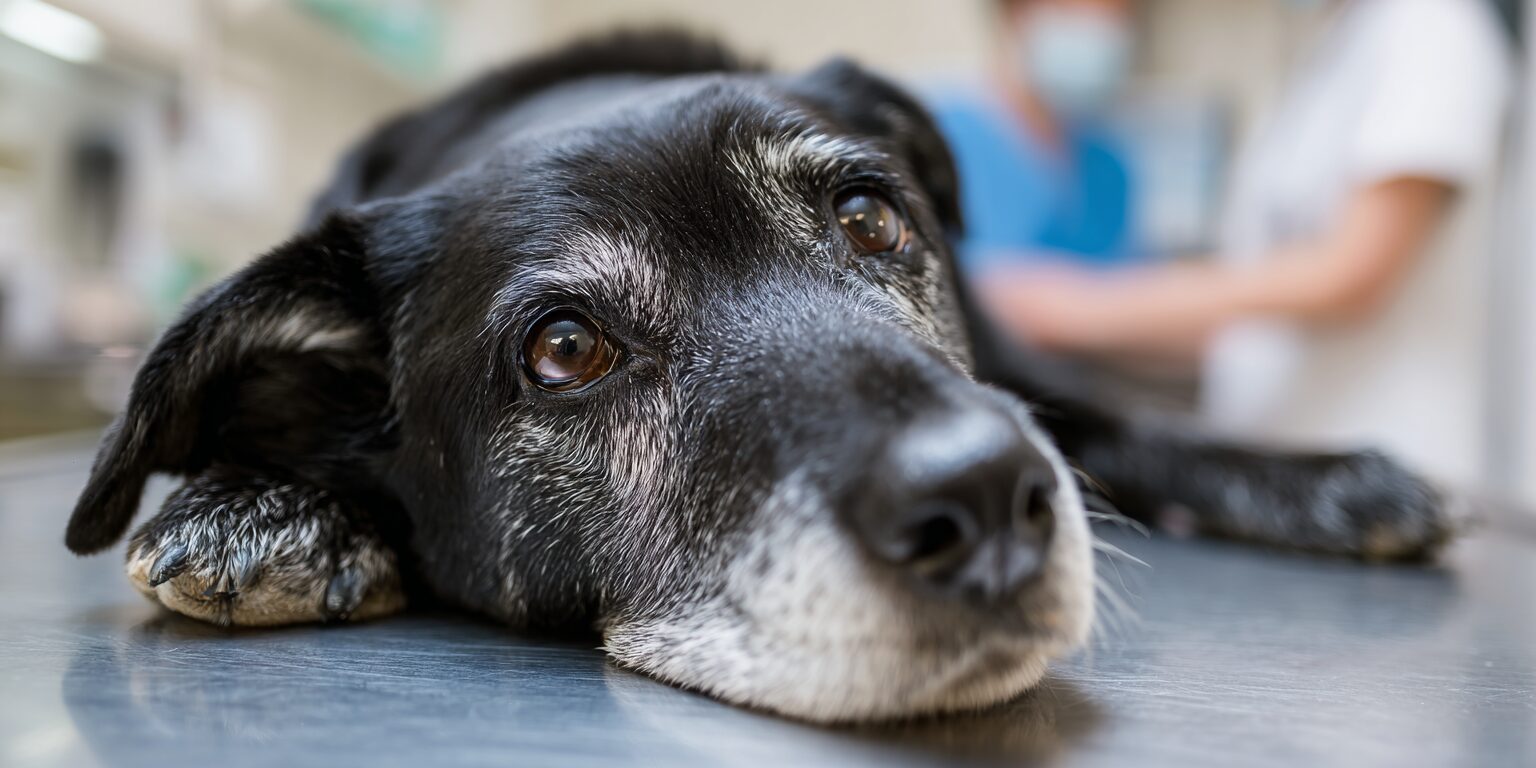
Diagnosing elbow dysplasia usually starts with a physical exam. Your veterinarian may watch your dog walk, check range of motion, feel the elbow joint, assess pain, and look for swelling or muscle loss.
Imaging is often needed to assess the condition of your dog’s joint.
Your veterinary team may recommend:
- X-rays
- CT scan
- Joint evaluation under sedation
- Referral to a surgical specialist
- Arthroscopic evaluation in selected cases
A CT scan can give a more detailed view of the canine elbow than standard x-rays, especially for fragmented coronoid process, elbow incongruity, and joint surface damage.
Most dogs are diagnosed with elbow dysplasia when they are young adults, often between 5 and 18 months of age, as this is when clinical signs typically become apparent.
Early detection matters because elbow dysplasia can lead to irreversible damage and progressive osteoarthritis. With proper management, many dogs can still have a good quality of life. Regular veterinary check-ups are essential for dogs with elbow dysplasia to monitor the progression of the condition and adjust treatment plans as necessary.
Treatment for elbow dysplasia in dogs
There is no true cure for elbow dysplasia, but treatment can relieve pain, improve function, and slow progression. While there is no cure for elbow dysplasia, treatment includes weight management, joint supplements, anti-inflammatory medications, and surgery when needed.
Treatment options depend on:
- Dog’s age
- Severity of joint damage
- Type of elbow abnormality
- Pain level
- Degree of arthritis
- Activity level
- Weight
- Whether one or both elbows are affected
Treatment for elbow dysplasia in dogs often includes a multimodal plan. That means using several tools together.
Medical management for mild cases
Mild cases may respond well to conservative care.
Medical management may include:
- Weight control
- Controlled exercise
- Exercise modification
- Joint supplements
- Pain medications
- Anti-inflammatory medications
- Physical therapy
- Red light therapy
- Regular veterinary check-ups
Weight management is huge. Extra weight increases force through the dog’s joint and can worsen joint pain, inflammation, and cartilage wear.
Joint supplements may support joint health over time. Pain relief medications and nonsteroidal anti-inflammatory drugs may help decrease pain and inflammation when prescribed by your veterinarian.
Regular check-ups help your veterinary team monitor progression and adjust the treatment plan when needed.
Physical therapy for elbow dysplasia
Physical therapy can be valuable for dogs with elbow dysplasia because it helps maintain joint function, reduce stiffness, and support muscle strength.
A rehab plan may include:
- Passive range-of-motion exercises
- Massage
- Controlled leash walks
- Strength exercises
- Balance work
- Hydrotherapy
- Low-impact movement
The goal is controlled movement without overloading the affected joint.
Too much activity can increase pain. Too little activity can lead to stiffness and muscle loss.
Balance matters.
Surgery for elbow dysplasia
Elbow dysplasia surgery may be recommended when there is significant joint damage, loose fragments, severe pain, or poor response to medical management. In advanced cases of elbow dysplasia, dogs may need to undergo surgery to restore joint function and relieve pain.
Surgical intervention is often necessary for advanced cases, with techniques including arthroscopy to remove damaged tissue and realignment procedures to improve joint function. Surgical treatment may include:
- Arthroscopic surgery
- Fragment removal
- Cartilage flap removal
- Proximal dynamic ulnar osteotomy
- Ulnar osteotomy
- Sliding humeral osteotomy
- Procedures to address medial compartment disease
- Elbow replacement in severe cases
Arthroscopic surgery is minimally invasive and may be used to remove damaged tissue, evaluate the joint surface, and treat a fragmented medial coronoid process or cartilage flap.
Proximal dynamic ulnar osteotomy may be used in some young dogs to change force through the elbow joint. Sliding humeral osteotomy may be considered for medial compartment disease by shifting weight away from the damaged medial aspect of the joint.
Total elbow replacement is a surgical option for severely degenerated joints. It is complex and carries risks of complications, it is not as commonly performed as hip replacements.
Your veterinary surgeon can explain which surgical procedures make sense for your dog’s elbow joint.
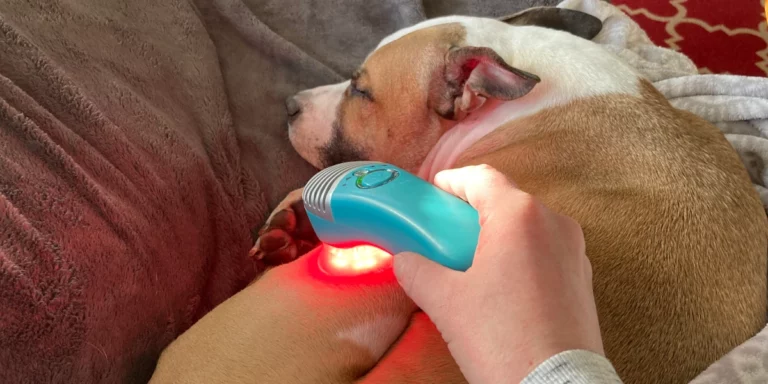
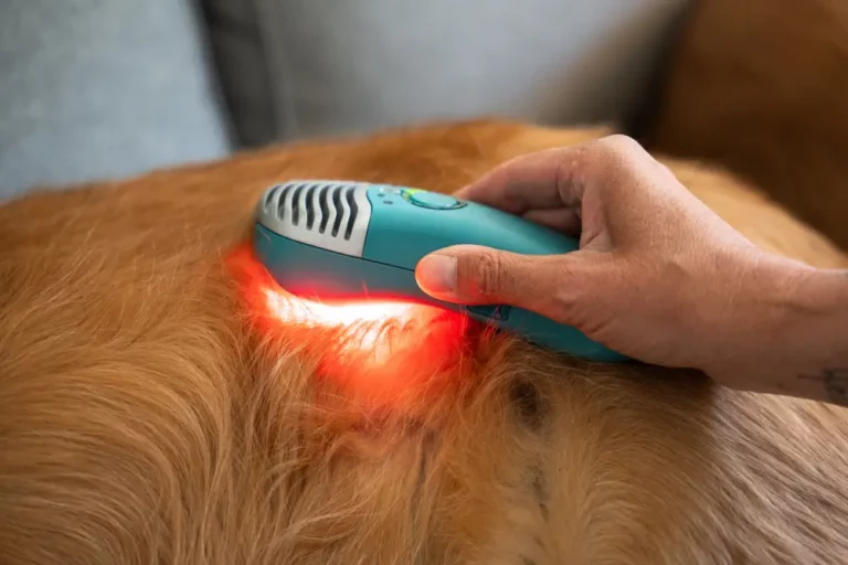
How red light therapy supports elbow dysplasia

Photobiomodulation, or PBM, also called red light therapy, low-level laser therapy, LED therapy, cold laser, and near-infrared therapy, refers to the same therapeutic category using light energy to influence cellular biology.
Red light therapy can help dogs with elbow dysplasia by supporting pain relief, reducing inflammation, improving local circulation, and helping the tissues around the affected joint recover after stress.
Learn more here: Science of Red Light Therapy.
Red light therapy does not correct abnormal bone growth or reverse joint incongruity. It can help manage the discomfort, inflammation, stiffness, and muscle tension that often come with elbow dysplasia.
For dogs recovering from elbow dysplasia surgery, red light therapy can support comfort, tissue repair, and inflammation control around the surgical area as part of the recovery plan.
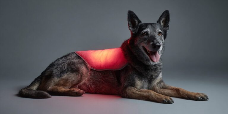
Why Luma is helpful for elbow dysplasia
The MedcoVet Luma gives pet owners a way to support their dog’s comfort at home.
Luma uses red and near-infrared light to support circulation, inflammation control, and tissue comfort around the dog’s elbow joint. It is non invasive, drug free, and built for pet parents to use with clinician guidance.
For elbow dysplasia, consistency matters.
Many dogs need long-term support. Luma can fit alongside weight management, physical therapy, joint supplements, medical management, and surgical recovery.
Luma may help support:
- Reduced joint discomfort
- Pain relief
- Lower inflammation
- Better comfort after walks
- Physical therapy tolerance
- Recovery after elbow dysplasia surgery
- Long-term joint health
- Comfort in dogs with chronic pain
Read more here: Red Light Therapy for Pets with Hip and Elbow Dysplasia.
For broader guidance, visit Red Light Therapy for Dogs.
Want to know if Luma fits your dog’s elbow dysplasia plan?
Take the quiz: Is red light therapy right for your dog?
Anderson
Real Dogs, Real Relief
Elbow dysplasia can make everyday movement harder. A walk gets shorter. A front-leg limp shows up after play. Stairs feel harder. Rest does not always look restful.
Luma gives pet parents a way to support comfort, mobility, and joint health at home, right where their dog feels safest.
Want to know if Luma could help your dog move more comfortably?
Hazel Moon
Can elbow dysplasia be prevented?
You cannot prevent every case of elbow dysplasia in dogs, especially when genetics are involved. However, you can take steps to help prevent elbow dysplasia by using genetic screening such as OFA health testing, choosing responsible breeders, and providing joint supplements to support joint health and reduce the risk or severity of the condition.
You can reduce risk.
Helpful steps include:
- Choose breeders who screen for elbow dysplasia
- Feed large-breed puppies correctly
- Avoid rapid weight gain
- Avoid overfeeding during growth
- Keep puppies lean
- Avoid repetitive high-impact exercise during growth
- Schedule veterinary exams if limping appears
- Keep adult dogs at a healthy weight
Preventive care starts early. Growing joints need steady nutrition, safe movement, and good breeding practices.
MedcoVet’s clinical view on elbow dysplasia
MedcoVet focuses on photobiomodulation protocols for dogs with elbow dysplasia, hip dysplasia, arthritis, surgery recovery, pain, and mobility issues.
Our clinical approach considers the affected joint, tissue depth, coat type, inflammation, pain pattern, surgery status, and long-term treatment plan. Elbow dysplasia is often lifelong. Dogs do best with a layered plan: veterinary diagnosis, weight control, pain management, physical therapy, joint supplements, and home support.
Red light therapy can be a useful part of that plan because it supports comfort, inflammation control, and day-to-day mobility.
Questions pet parents ask about elbow dysplasia in dogs
Clinical summary
Mechanism:
Elbow dysplasia is a developmental orthopedic condition involving abnormal fit, growth, or cartilage development in the canine elbow. The elbow joint includes the humerus, radius, and ulna. Joint incongruity, fragmented medial coronoid process, ununited anconeal process, osteochondrosis, and medial compartment disease can damage the joint surface and lead to degenerative joint disease.
Evidence level:
Diagnosis and treatment of canine elbow dysplasia are well established in veterinary medicine. X-rays, CT scan, and arthroscopy are commonly used diagnostic tools. Weight control, exercise modification, pain medications, joint supplements, physical therapy, and surgical management are standard treatment options. Photobiomodulation has moderate support for pain relief, inflammation control, and recovery support.
When red light therapy works best:
Red light therapy works best when elbow dysplasia causes joint pain, inflammation, stiffness, reduced mobility, or discomfort during rehab. It may also support comfort after elbow dysplasia surgery as part of a veterinarian-guided recovery plan.
When not to use red light therapy:
Do not use red light therapy over a known or suspected tumor, untreated infection, or open wound without veterinary guidance. Dogs with severe sudden lameness, swelling, trauma, or rapid decline need veterinary care.
Help your dog move with less elbow pain
Elbow dysplasia in dogs can affect movement, play, comfort, and long-term joint health.
You have options.
Prefer to talk through your pet’s condition with a clinician?
Dave
Evidence Citations

About the Author
Alon Landa is the CEO and co-founder of MedcoVet, a leader in at-home red light therapy for pets. With over 20 years of experience in medical technology and firsthand involvement in developing the Luma, Alon combines deep technical knowledge with a passion for improving pet health. He regularly collaborates with veterinarians and pet parents to advance photobiomodulation (PBM) care at home.
📍 Based in Boston, MA
📖Read more from Alon here

About the Medical Reviewer
Clinical Focus: Surgery, anesthesia, canine fitness, injury prevention, agility
Kristy Williams brings over 30 years of experience to the veterinary field. Her career began in the 1990s, working as a civilian for the Army Veterinary Corps at RAF Feltwell in England, where she first discovered her passion for animal care and supporting their families. Upon returning to the United States, Kristy pursued her education and graduated in 2005 as a certified veterinary technician after passing the national exam. She has since gained extensive experience in both general practice and emergency/referral practices.
Read More about Kristy here.
🐾 Ready to Take the Next Step?
Whether you’re just learning or ready to act — we’ve got you.
👉 Book a Free Consult
Talk to a licensed clinician about your pet’s condition and get a personalized plan. No pressure, just real help.
👉Learn More About the Luma
Explore how our at-home red light therapy device works, why it’s different, and what it can do for your pet.