Signs of Hip Dysplasia in Dogs
How to spot early signs, when to call your vet, and how to support comfort and mobility
Noticing changes in your dog’s hips or back legs?
Talk with a MedcoVet clinician about your dog’s symptoms, mobility, pain level, and next steps.
Medically reviewed by: Kristy Williams, CVT, CCFT (Specialties: Pet rehabilitation, pain management, photobiomodulation)
Reviewed: June 2026
Updated: June 2026
What are the signs of hip dysplasia in dogs?
The most common clinical signs of hip dysplasia in dogs include difficulty rising, limping in the hind legs, a bunny hopping gait, stiffness, reluctance to run or jump, pain around the hip joint, and chronic or occasional lameness. These clinical signs may also present as limping without previous trauma, difficulty standing up, and decreased activity levels—such as a significant drop in enthusiasm for walking, playing, or jumping into cars. Some dogs show early signs as young as 4 months old, while others develop symptoms later as arthritis and joint damage progress. If your dog is showing changes in mobility, gait, or comfort, a veterinary exam and x rays can help confirm whether hip dysplasia is the cause.
If your dog is already showing signs of pain, stiffness, or reduced mobility:
Want help understanding your dog’s symptoms?
Sprite
What is hip dysplasia in dogs?
Hip dysplasia is a common skeletal condition where the dog’s hip joint does not form correctly. The hip is a ball and socket joint. The femoral head, which is the ball at the top of the thigh bone (femur), should fit smoothly into the hip socket in the pelvic bone.
With canine hip dysplasia, the ball and socket joint is loose, shallow, or poorly aligned. That poor fit creates joint instability. Over time, the hip joint can become inflamed, painful, and damaged.
Many dogs with hip dysplasia develop arthritis, bone spurs, stiffness, muscle loss, and reduced range of motion. The condition can affect one or both hips.
Hip dysplasia in dogs can look mild at first. A little hesitation. A slower start in the morning. A skipped jump into the car.
Then it becomes harder to ignore.
Early signs of hip dysplasia

Early diagnosis matters because treatment can begin before joint damage becomes more advanced.
Watch for these early signs:
- Difficulty rising after rest
- Stiffness after naps
- Limping without previous trauma
- Occasional hind end lameness
- Reluctance to run, jump, or climb stairs
- Bunny hopping when moving quickly
- Trouble getting on and off furniture
- Abnormal sitting positions
- Reduced range of motion in the hips
- Less enthusiasm for walks or play
- Pain when the hips are touched
- Muscle atrophy in the hind legs
A bunny hopping gait means the dog moves both hind legs together instead of using them independently. Pet parents often notice this when the dog runs, climbs stairs, or moves uphill.
Hind end lameness may be persistent or occasional. Some affected dogs limp every day. Others look fine one day, then stiff or sore the next.
Some veterinarians take advantage of the anesthesia during spaying or neutering to perform pelvic X-rays. This allows for clearer imaging, which can help diagnose hip dysplasia early, even before symptoms appear, enabling timely treatment options.
Later signs of hip dysplasia
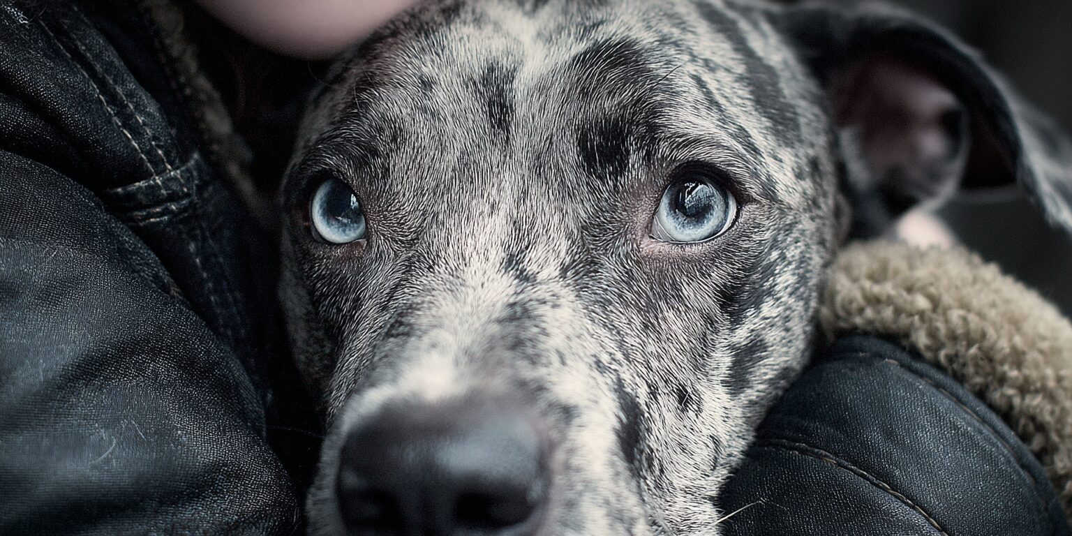
As hip dysplasia progresses, dogs may show clearer signs of pain and joint damage.
These can include:
- Trouble standing up from lying down
- Avoiding stairs
- Refusing to jump into the car
- Reduced activity
- Visible pain in the hip area
- Worsening lameness after exercise
- Loss of muscle mass in the hind limbs
- Bigger shoulder muscles from shifting weight forward
- Clicking or popping in the hip joint
- Irritability when handled
- Less interest in normal routines
Thigh muscle atrophy is common in dogs with hip dysplasia. The hind legs may look smaller because the dog avoids using them fully. At the same time, the shoulder muscles may become more developed because the dog shifts weight onto the front end.
That weight shift protects the hips, but it can create stress in other areas of the body.
Which dogs are most at risk?
Genetics plays a major role in developing hip dysplasia. Large breed dogs and giant breed dogs have a higher risk, especially breeds such as German Shepherds, Labrador Retrievers, Golden Retrievers, Great Danes, Rottweilers, and Saint Bernards.
Large breed puppies are especially vulnerable during growth.
Risk factors include:
- Genetic predisposition
- Rapid growth
- Excessive exercise during the growth phase
- Improper nutrition
- Excess weight gain
- Poor muscle development
- Joint stress from high-impact activity
Environmental factors matter too. Improper nutrition, unbalanced diets, and fast weight gain can increase a dog’s risk of developing hip dysplasia. Excessive exercise in young dogs can place undue stress on developing joints.
Healthy hips start early.
If you are getting a puppy from a breeder, ask whether the breeding dogs were screened for hip dysplasia. To help prevent hip dysplasia, early screening, maintaining a healthy weight, and providing proper nutrition are important steps. For large-breed puppies, feeding a diet formulated for large-breed growth until at least 12 to 18 months of age can help support proper nutrition and joint development.
How veterinarians diagnose hip dysplasia
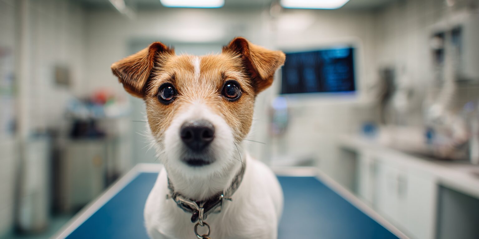
Diagnosing hip dysplasia usually begins with a physical exam. Your veterinarian may watch your dog walk, feel the dog’s hips, assess range of motion, check for pain, look for muscle atrophy, and check for signs of discomfort or abnormal joint movement in the dog’s hips.
X rays (radiographs) are commonly used to confirm hip dysplasia diagnosed by exam and to assess the severity of changes in the dog’s hip joint, including the hip socket, femoral head, joint laxity, arthritis, and bone changes.
Your veterinarian may also ask about:
- When symptoms started
- Whether the signs are getting worse
- Your dog’s age and breed
- Exercise habits
- Weight history
- Diet and supplements
- Prior injuries
- Response to pain medications
A clear diagnosis helps guide the treatment plan.
How hip dysplasia is treated
Managing hip dysplasia early is crucial for improving outcomes and supporting your pet’s health and mobility. Hip dysplasia treatment plans depend on the dog’s age, severity, pain level, activity, and whether medical or financial reasons affect available options.
Non-surgical treatment options may include:
- Weight management (including weight loss)
- Low impact exercise
- Physical therapy
- Physical rehabilitation
- Joint supplements
- Pain medications
- Nonsteroidal anti inflammatory drugs
- Joint fluid modifiers
- Anti inflammatory medications
- Lifestyle changes
- Home modifications
- Red light therapy
Hips exercise restriction is also important—limiting your dog’s activity on hard surfaces can help reduce joint stress and prevent the condition from worsening.
Medical management often uses a multimodal approach. That means combining several tools to alleviate pain, support joint health, preserve muscle mass, and improve the dog’s quality of life while maintaining your pet’s mobility and overall health.
Joint supplements such as glucosamine and chondroitin may be recommended to support joint health. Your veterinarian can help you decide when to start joint supplements, especially if your dog has a high risk of developing hip dysplasia.
Non-surgical treatments for hip dysplasia may involve weight management, physical therapy, and the use of joint supplements like glucosamine and chondroitin to support joint health.
Weight management is one of the most important pieces. A healthy weight and weight loss reduce strain on the hip joint and help affected dogs move more comfortably.
By managing hip dysplasia with early detection and a tailored treatment plan, you can directly influence your dog’s quality of life and help them stay active and comfortable for years to come.
Surgical options for hip dysplasia
Some dogs need surgical intervention, especially when pain is severe, mobility is poor, or conservative care is not enough. Surgical procedures are designed to modify or replace the dog’s hip joint to improve mobility and alleviate pain.
Several surgical procedures may be considered, including:
- Total hip replacement
- Femoral head ostectomy
- Triple pelvic osteotomy
These surgical options can significantly improve mobility and reduce pain in affected dogs.
Total hip replacement replaces the damaged hip joint with an artificial joint. It can significantly improve mobility and reduce pain in the right candidate.
Femoral head ostectomy removes the femoral head, allowing the body to form a false joint with scar tissue. It is often considered for smaller dogs, though some larger dogs may also be candidates.
Triple pelvic osteotomy is generally performed in young dogs before severe arthritis develops. It changes the position of the pelvic bone to improve hip socket coverage.
Your veterinary surgeon can explain which option best fits your dog’s condition.
How red light therapy supports dogs with hip dysplasia

Photobiomodulation, or PBM, also called red light therapy, low-level laser therapy, LED therapy, cold laser, and near-infrared therapy, refers to the same therapeutic category using light energy to influence cellular biology.
Red light therapy can help dogs with hip dysplasia by reducing inflammation, supporting blood flow, and helping calm pain signals. It may also support muscle comfort and mobility when used as part of a comprehensive treatment plan.
Learn more here: Science of Red Light Therapy.
Red light therapy does not reverse structural hip changes. It can help manage the inflammation, pain, arthritis, and muscle strain that often come with hip dysplasia.
For dogs with stiffness, difficulty rising, and reduced mobility, that support can make daily life feel easier.
Why Luma is helpful for hip dysplasia
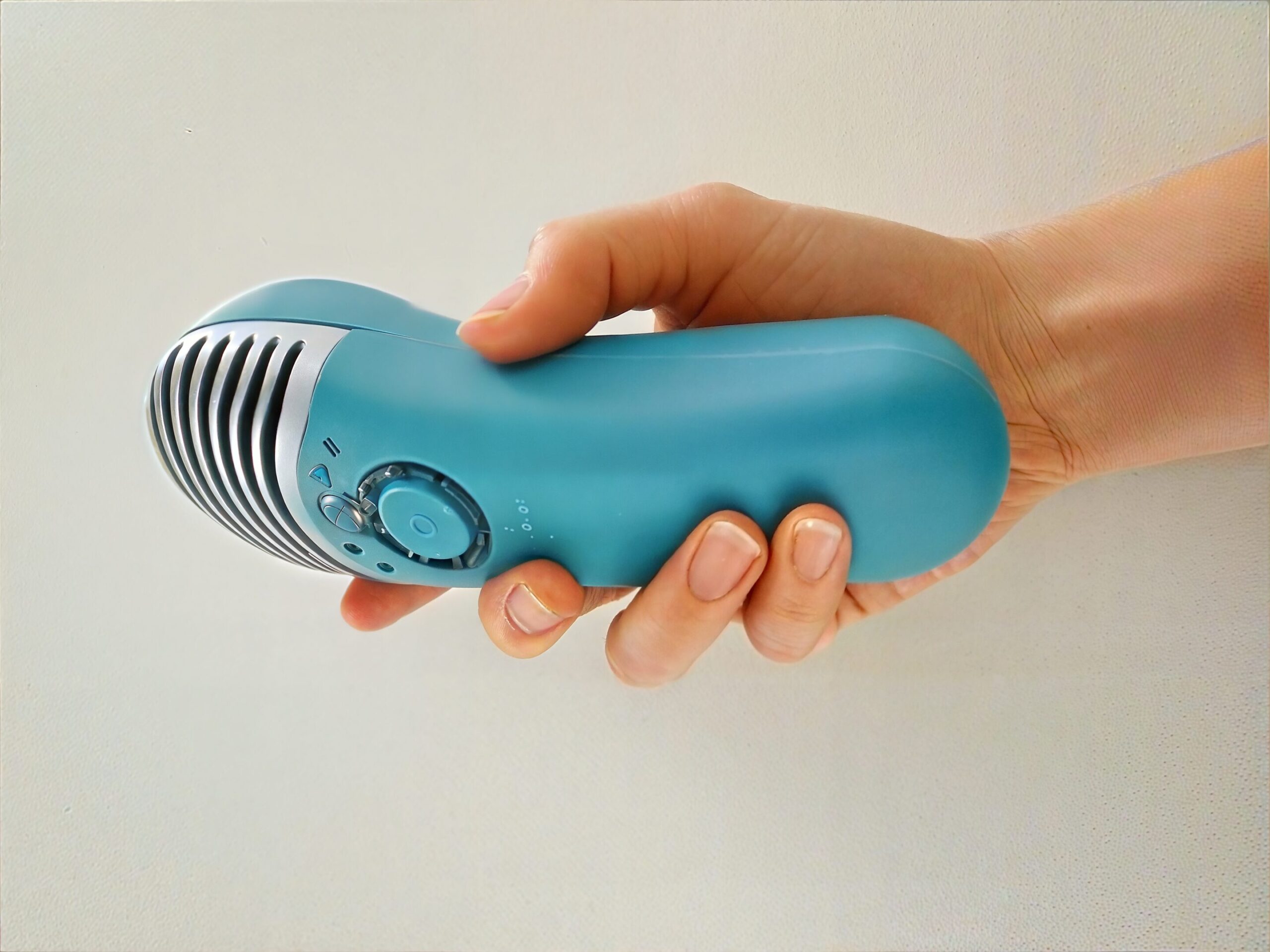
The MedcoVet Luma gives pet parents a way to support joint comfort at home.
Luma uses red and near infrared light to support pain relief, circulation, inflammation control, and tissue recovery around the affected area. It is non invasive, drug free, and designed for home use with clinician guidance.
That matters for chronic conditions.
Dogs with hip dysplasia often need long term support, not a one-time treatment. Luma can fit into a broader treatment plan with physical therapy, low impact exercise, weight management, joint supplements, and veterinary care.
Luma may help support:
- Reduced hip pain
- Improved mobility
- Better comfort after exercise
- Less stiffness after rest
- More willingness to move
- Muscle comfort around the dog’s hips
- Long term joint health
Read more here: Red Light Therapy for Pets with Hip and Elbow Dysplasia.
For broader guidance, visit Red Light Therapy for Dogs.
👉 Want to know if Luma is right for your dog’s hip dysplasia signs?
Gideon
Real Stories from Dogs Using Luma
Every dog with hip dysplasia moves a little differently. Some struggle to stand. Some avoid stairs. Some still want to play, but their hips make it harder.
These stories show what can happen when pet parents add Luma to their dog’s comfort and mobility plan.
Want to know if Luma could help your dog move more comfortably?
Hazel Moon
MedcoVet’s clinical view on hip dysplasia support
MedcoVet focuses on photobiomodulation protocols for dogs with pain, arthritis, surgery recovery, hip dysplasia, elbow dysplasia, and mobility issues.
Our clinical approach considers tissue depth, coat type, treatment location, activity level, pain pattern, and the dog’s overall treatment plan. Hip dysplasia in dogs often involves more than the hip joint. It can affect the hind limbs, shoulder muscles, gait, activity, confidence, and the pet’s mobility.
The strongest plans usually combine veterinary diagnosis, healthy weight, proper nutrition, low impact exercise, physical therapy, pain management, joint supplements, and at-home support.
Questions pet parents ask about hip dysplasia signs
Clinical summary
Mechanism:
Hip dysplasia is a developmental orthopedic condition in which the femoral head and hip socket do not fit correctly. Joint instability leads to inflammation, cartilage wear, arthritis, pain, and compensatory muscle changes. Red light therapy supports cellular activity, blood flow, and inflammatory balance around affected tissues.
Evidence level:
Hip dysplasia diagnosis and treatment are well established in veterinary medicine. X rays are the standard imaging tool. Weight management, pain medications, physical therapy, joint supplements, and surgery are common treatment options. Photobiomodulation has moderate support for pain relief, inflammation reduction, and mobility support in musculoskeletal conditions.
When it works best:
Red light therapy works best when dogs have pain, inflammation, stiffness, secondary arthritis, or muscle strain related to hip dysplasia. It is most useful as part of a comprehensive treatment plan that includes veterinary diagnosis, weight control, low impact exercise, and joint support.
When not to use:
Do not use red light therapy over a known or suspected tumor, untreated infection, or unexplained pain without veterinary guidance. Dogs with sudden lameness, severe pain, or rapidly worsening mobility should be evaluated by a veterinarian.
Help your dog move with less pain
Signs of hip dysplasia in dogs can start small, but they matter. Difficulty rising, bunny hopping, reluctance to climb stairs, and changes in the hind legs are all worth taking seriously.
Early diagnosis gives your dog more options.
If your dog is showing signs of hip dysplasia and you want to know whether red light therapy may help:
Want help thinking through your dog’s symptoms and next steps?
Dave
Dave
Evidence Citations
🐾 Ready to Take the Next Step?
Whether you’re just learning or ready to act — we’ve got you.
👉 Book a Free Consult
Talk to a licensed clinician about your pet’s condition and get a personalized plan. No pressure, just real help.
👉Learn More About the Luma
Explore how our at-home red light therapy device works, why it’s different, and what it can do for your pet.

About the Author
Alon Landa is the CEO and co-founder of MedcoVet, a leader in at-home red light therapy for pets. With over 20 years of experience in medical technology and firsthand involvement in developing the Luma, Alon combines deep technical knowledge with a passion for improving pet health. He regularly collaborates with veterinarians and pet parents to advance photobiomodulation (PBM) care at home.
📍 Based in Boston, MA
📖Read more from Alon here

About the Medical Reviewer
Clinical Focus: Surgery, anesthesia, canine fitness, injury prevention, agility
Kristy Williams brings over 30 years of experience to the veterinary field. Her career began in the 1990s, working as a civilian for the Army Veterinary Corps at RAF Feltwell in England, where she first discovered her passion for animal care and supporting their families. Upon returning to the United States, Kristy pursued her education and graduated in 2005 as a certified veterinary technician after passing the national exam. She has since gained extensive experience in both general practice and emergency/referral practices.
Read More about Kristy here.